AQA A-Level Biology: The Immune System
A clear revision guide to the immune system for AQA A-Level Biology: antigens, phagocytosis, T and B lymphocytes, antibodies, vaccines, HIV, and monoclonal antibodies.
The immune system tells the body's own cells apart from anything foreign, and destroys what does not belong. This guide covers how cells are recognised, how the two arms of the specific immune response work, and how this understanding is used in vaccines, HIV research and monoclonal antibodies.
Antigens and cell recognition
An antigen is a foreign molecule, usually a protein, glycoprotein or glycolipid, that triggers an immune response, including the production of antibodies.
Every cell carries specific molecules on its surface that identify it. Because these are often proteins with a particular tertiary structure, the immune system can use them like a label. This lets it identify pathogens such as bacteria, viruses and fungi, cells from another organism of the same species (as in a transplant), abnormal cells such as cancer cells or virus-infected cells, and toxins released by bacteria.
Phagocytosis: the non-specific response
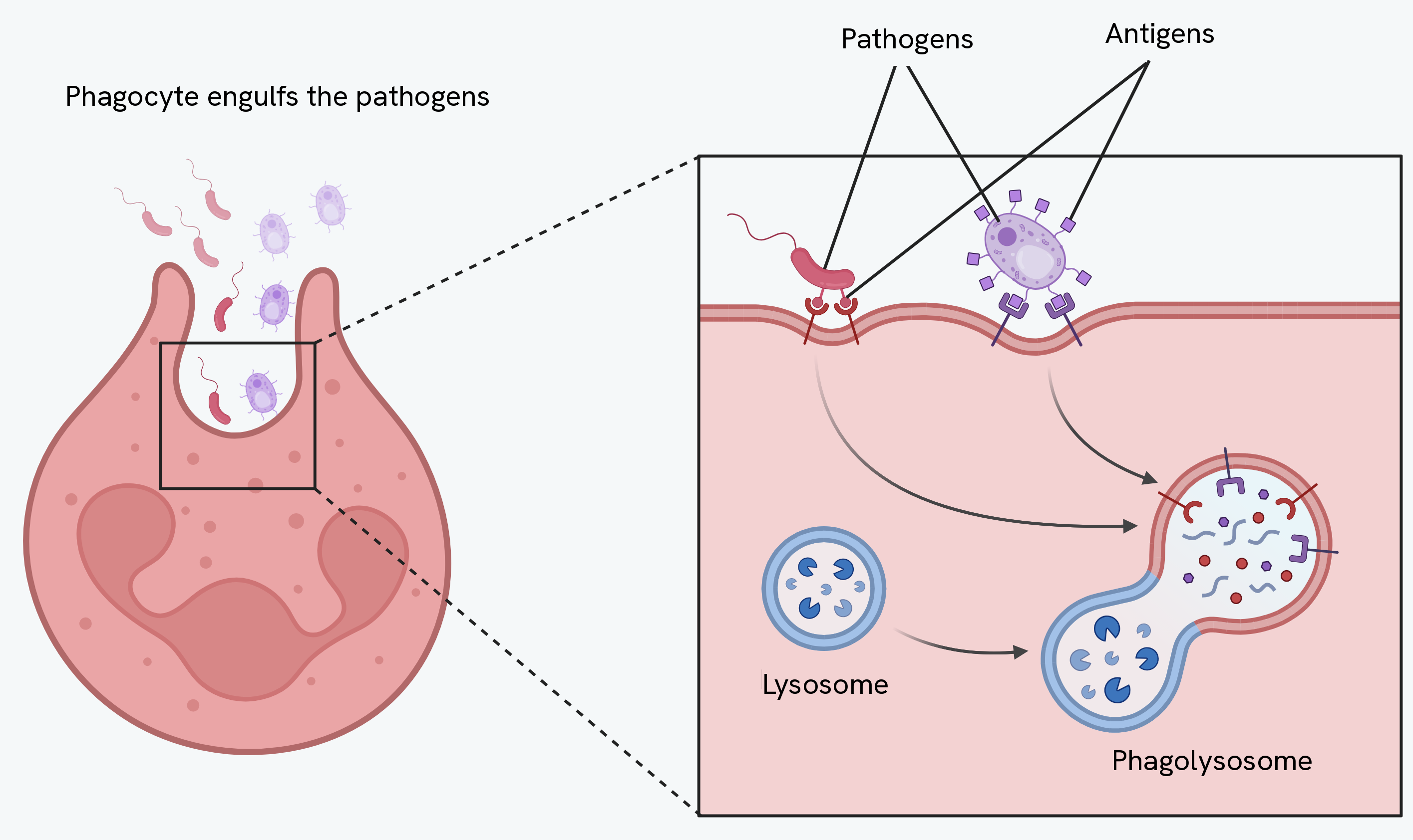
The first response to a pathogen does not depend on which pathogen it is. A white blood cell called a phagocyte is attracted to the pathogen and recognises its foreign antigens. The phagocyte engulfs the pathogen by surrounding it with its membrane, trapping it in a vesicle called a phagosome. A lysosome then fuses with the phagosome and releases hydrolytic enzymes (lysozymes), which digest the pathogen.
Phagocytosis also sets up the specific response: the phagocyte displays the pathogen's antigens on its own surface, becoming an antigen-presenting cell.
The cellular response: T lymphocytes
T lymphocytes respond to antigens that are being presented on the surface of another cell, such as an infected body cell or a phagocyte.
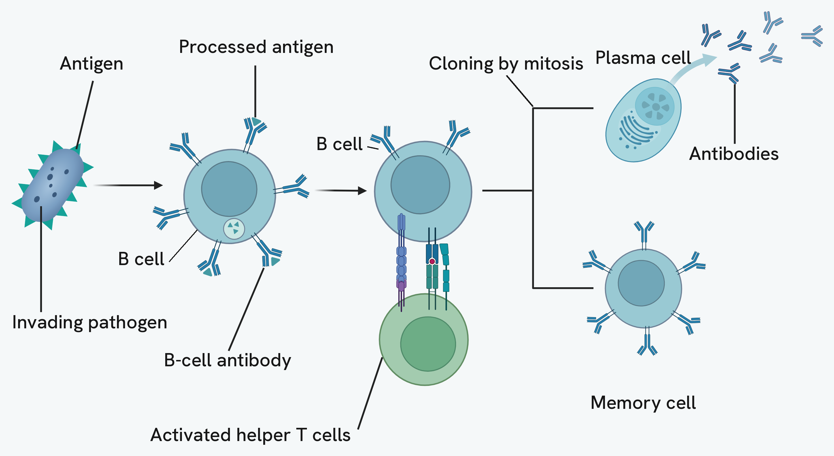
A helper T cell with a receptor complementary to the presented antigen binds to it. This activates the helper T cell, which then divides by mitosis to form clones. These clones stimulate the rest of the response: they activate cytotoxic T cells, which kill infected and abnormal cells (by releasing a protein called perforin), they stimulate B lymphocytes, and they stimulate more phagocytes.
The humoral response: B lymphocytes
B lymphocytes respond to antigens directly. Each B cell carries a different antibody on its surface. When a B cell's antibody meets a complementary antigen, that particular cell is selected, a process called clonal selection, and helper T cells stimulate it to divide rapidly by mitosis into clones.
These clones become two kinds of cell. Plasma cells secrete large quantities of the specific antibody into the blood, dealing with the current infection. Memory cells remain in the blood long after the infection has cleared, ready to respond quickly if the same antigen returns.
Antibodies
An antibody is a protein with a quaternary structure, made of four polypeptide chains held together by disulfide bridges. Each antibody has a variable region whose specific tertiary structure forms a binding site complementary to one particular antigen, and a constant region that is the same across antibodies. A flexible hinge region lets the two binding sites reach antigens that are different distances apart.
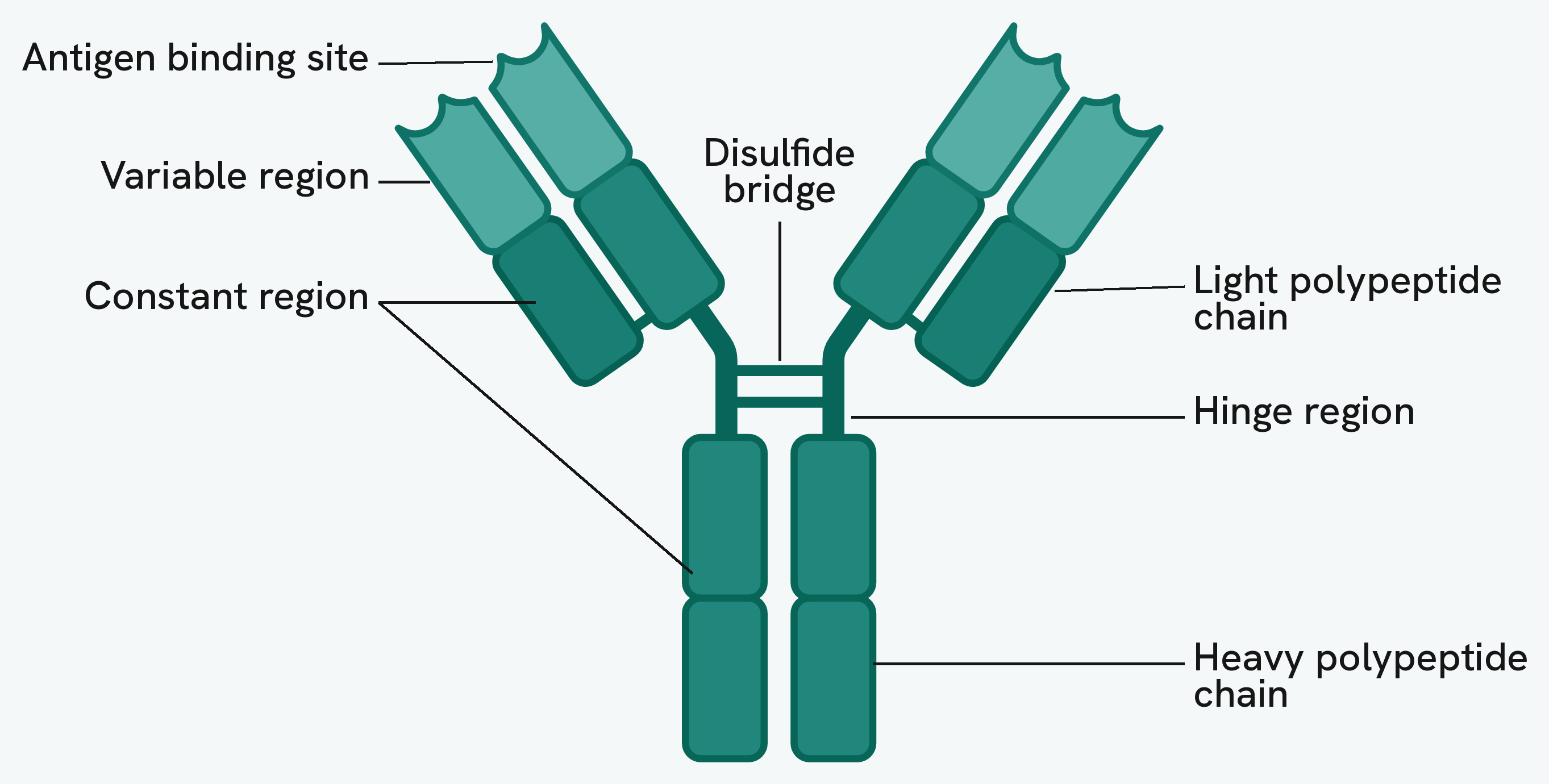
When an antibody meets its antigen it binds to form an antigen-antibody complex. Because each antibody has two binding sites, it can bind two pathogens at once, clumping them together in a process called agglutination. This makes the pathogens easier for phagocytes to engulf several at a time, and antibodies also attract phagocytes to the site.
The primary and secondary response
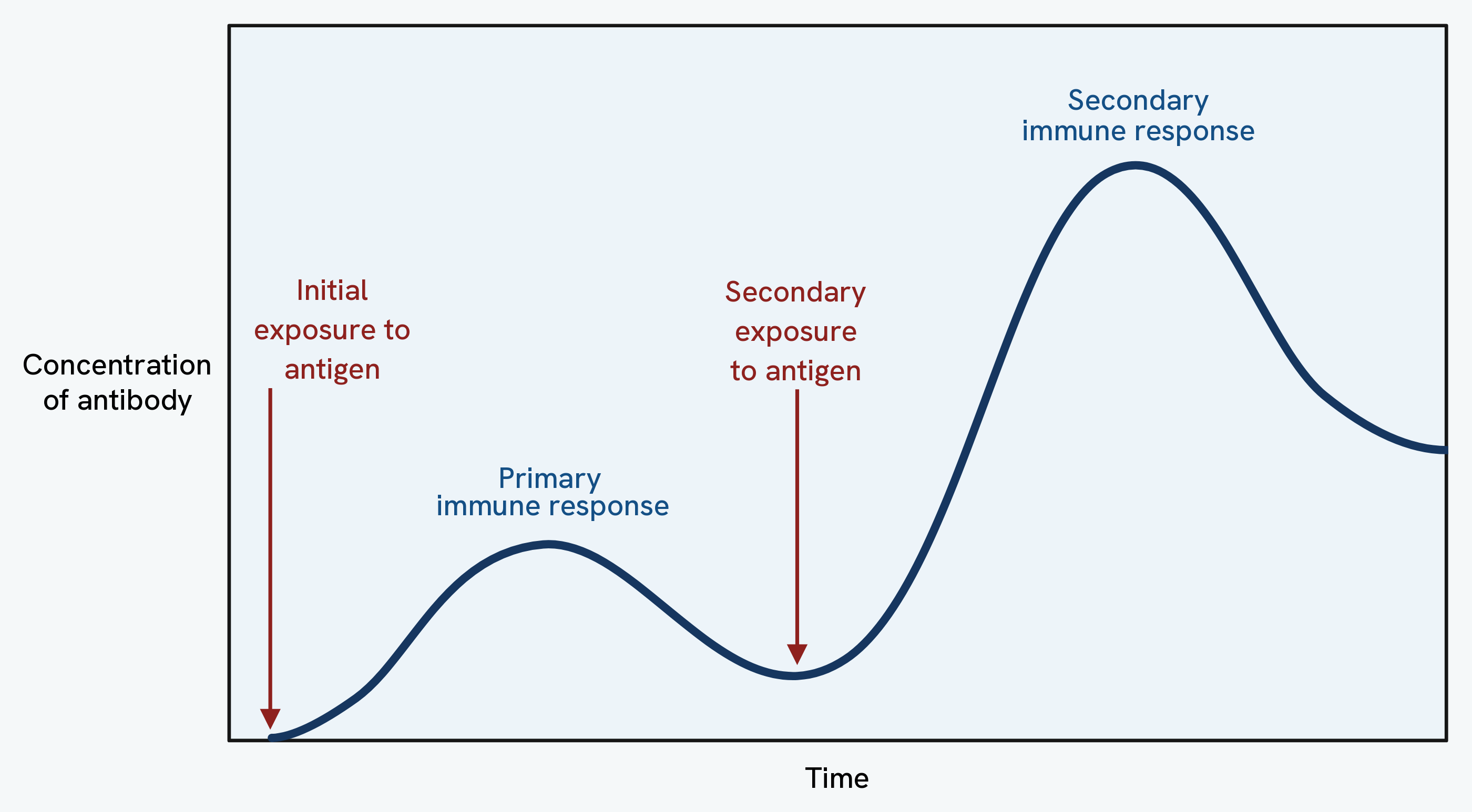
The first time the body meets an antigen, the primary response is slow: it takes time for the right B cell to be selected and for plasma cells to build up, so antibodies are produced slowly and at a low concentration. Crucially, this first exposure also produces memory cells.
If the same antigen is met again, the secondary response is much faster and stronger. The memory cells divide rapidly into plasma cells, so antibodies are produced quickly and at a much higher concentration, usually destroying the pathogen before it can cause symptoms. This is the basis of long-term immunity.
Vaccination
A vaccine introduces antigens into the body, often from dead or weakened (attenuated) pathogens, without causing the disease. This triggers a primary response and, importantly, the production of memory cells. If the real pathogen is met later, the memory cells produce a fast, strong secondary response, so the person does not become ill.
Vaccination also protects populations through herd immunity. When a large enough proportion of a population is immune, there are too few people for the pathogen to spread through, so even unvaccinated people are much less likely to catch it.
Active and passive immunity
Immunity can be gained in two ways, and they differ in how long they last.
| Feature | Active immunity | Passive immunity |
|---|---|---|
| How it arises | The body meets an antigen (infection or vaccine) | Antibodies are given from another organism |
| Memory cells | Produced | Not produced |
| Antibody source | Made by the body's own plasma cells | Introduced ready-made, for example in breast milk or across the placenta |
| Speed | Slower to develop | Fast-acting |
| Duration | Long-lasting | Short-lived, as the antibodies are broken down |
Antigenic variability
Some pathogens change over time. A mutation can change the shape of a pathogen's antigens, creating a new strain. When this happens, the memory cells and antibodies from a previous infection or vaccine no longer fit the changed antigen, so the person is no longer immune. This antigenic variability is why a new flu vaccine is needed each year, why there is still no effective vaccine for HIV, and why you can catch a cold many times.
HIV and AIDS
HIV is a virus with a lipid envelope, attachment proteins, a capsid, two strands of RNA and an enzyme called reverse transcriptase.
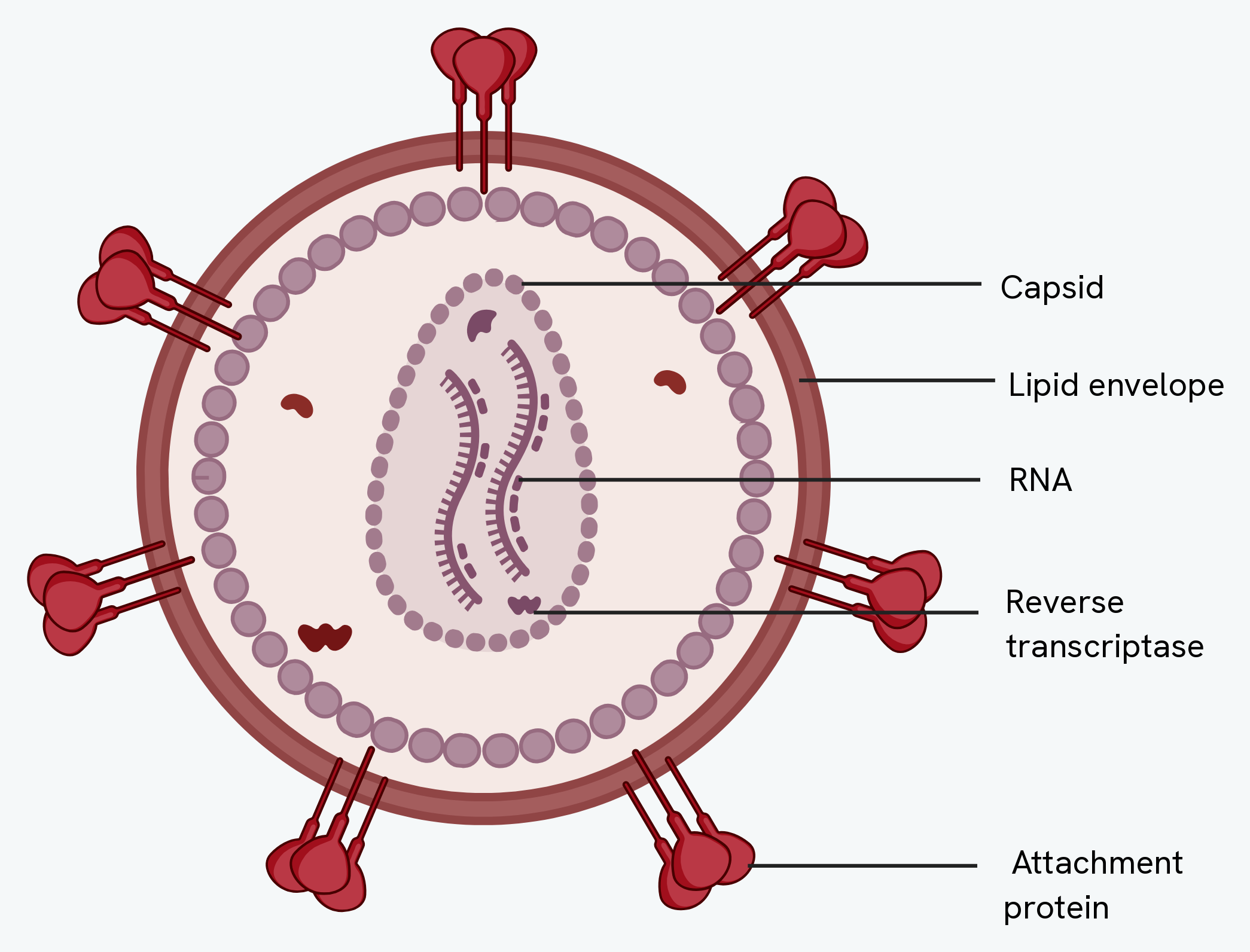
HIV infects helper T cells. Its attachment proteins bind to receptors on the helper T cell, and the lipid envelope fuses with the cell membrane, releasing the capsid inside. The capsid uncoats to release the RNA and reverse transcriptase, which converts the viral RNA into DNA. This viral DNA is inserted into the host cell's own DNA, where it can lie dormant. When it is active, it is transcribed and translated to make new viral proteins, and new virus particles are assembled and released to infect more cells.
Because HIV destroys helper T cells, and those cells are needed to stimulate cytotoxic T cells, B cells and phagocytes, the whole immune system is weakened. This is AIDS: with too few helper T cells, the body cannot fight off infections that it would normally cope with, and a person becomes vulnerable to opportunistic infections.
This also explains why antibiotics do not work on viruses. Antibiotics target features of bacteria, such as their murein cell wall and their metabolic processes. Viruses have neither their own metabolism nor a bacterial cell wall, and they hide inside host cells, so there is nothing for an antibiotic to attack.
Monoclonal antibodies
A monoclonal antibody is an antibody produced from a single clone of identical B cells, so every molecule has the same tertiary structure and binds the same target. This specificity makes monoclonal antibodies powerful tools in medicine.
In treatment, a monoclonal antibody can be made complementary to a molecule found only on a particular cell type, such as a cancer cell. A drug is attached to the antibody, which then binds to the target cell and delivers the drug precisely there, sparing healthy cells. Some monoclonal antibodies work simply by binding to and blocking a receptor or antigen.
In diagnosis, a monoclonal antibody is made complementary to a molecule linked to a condition, and a marker such as a dye or fluorescent tag is attached, so that when the antibody binds its target the location can be seen.
The ELISA test
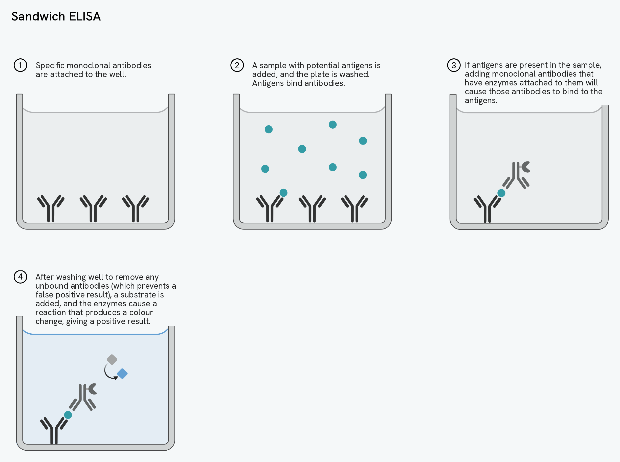
The ELISA test uses antibodies and a colour change to detect whether a particular antigen or antibody is present in a sample.
In one common form, a sample is fixed to a well and a monoclonal antibody with an enzyme attached is added, which binds if the target antigen is present. The well is washed to remove any unbound antibody, and then a substrate is added. If the enzyme-linked antibody is still there, the enzyme converts the substrate and produces a colour change, a positive result. A version that detects antibodies rather than antigens works the same way, but starts by fixing a known antigen to the well.
The washing step is essential: if unbound antibody is left behind, its enzyme will still produce a colour change and give a false positive. A control well is used to show that the colour change is caused only by the target being present, and not by anything else.
Judging the evidence
Because vaccines and monoclonal antibodies are used on large numbers of people, the evidence behind them has to be examined carefully. There are ethical questions, such as the use of animals in testing and the risk of side effects to people in clinical trials, weighed against the benefit of reducing suffering. There are also questions about methodology: whether the sample was large and diverse enough to be representative, whether control or placebo groups were used, whether trials ran long enough to reveal long-term effects, and whether they were double-blind to remove bias. Reading results critically, looking at how often side effects occurred and whether differences were statistically significant, is part of using these treatments responsibly.