AQA A-Level Biology: Microscopy and Studying Cells
A clear revision guide to studying cells for AQA A-Level Biology: magnification and resolution, optical, TEM and SEM microscopes, magnification calculations and cell fractionation.
We know what is inside cells because of the tools used to look at them. This guide covers how microscopes work, the difference between magnification and resolution, how to handle magnification calculations, and how cell fractionation separates organelles so they can be studied.
Magnification and resolution
These two words are often confused, but they mean different things.
Magnification is how many times larger an image is than the real object. It is calculated as:
- magnification = size of image divided by size of the real object
Resolution is the smallest distance between two points at which they can still be told apart as two separate points. It sets the limit on how much detail you can actually see. Turning the magnification up beyond the resolution just gives a bigger blurry image, not more detail.
Resolution is limited by the wavelength of whatever is used to view the specimen. Light has a relatively long wavelength, so an optical microscope cannot resolve anything closer together than about 200 nm. Electrons have a much shorter wavelength, which is why electron microscopes reveal far more detail.
Types of microscope
There are three microscopes to compare, and each involves a trade-off.
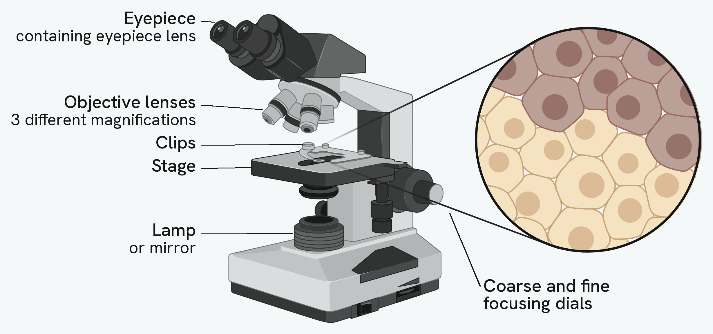
An optical (light) microscope focuses light through glass lenses and onto the specimen. Different structures absorb different amounts and wavelengths of light, producing a two-dimensional image of a cross-section. Its resolution is low because of the long wavelength of light, so it cannot show the internal structure of organelles. Its advantages are that preparation is simple, specimens can be alive, and images can be in colour.
A transmission electron microscope (TEM) passes a beam of electrons through a very thin specimen, focused by electromagnets. Denser parts absorb more electrons and appear darker, giving a two-dimensional image. Because electrons have such a short wavelength, the resolution is very high and internal structures such as the inside of organelles can be seen. The drawbacks are that the specimen must be dead and dehydrated, because the microscope uses a vacuum, and the complex preparation can introduce artefacts.
A scanning electron microscope (SEM) also uses electrons focused by electromagnets, but here the electrons bounce off the surface of the specimen rather than passing through it. This builds a three-dimensional image of the surface. The specimen does not need to be thin, but as with the TEM it must be dead, and the images are not in colour.
| Feature | Optical | TEM | SEM |
|---|---|---|---|
| Uses | Light through glass lenses | Electrons through the specimen | Electrons off the surface |
| Resolution | Low (about 200 nm) | Very high (about 0.2 nm) | High (about 0.2 nm) |
| Image | 2D cross-section | 2D cross-section, internal detail | 3D surface |
| Living specimens | Yes | No | No |
| Colour | Yes | No | No |
Artefacts
An artefact is a structure that appears in an image because of how the specimen was prepared, not because it was really there in the living cell. Because electron microscopy needs such heavy preparation, artefacts are a real risk. Scientists could tell artefacts from genuine organelles by preparing specimens in different ways: a structure that showed up with one technique but not another was more likely to be an artefact, while something seen consistently across techniques was more likely to be real.
Magnification calculations
Magnification, real size and image size are linked by one relationship, often remembered as I = A x M: image size equals actual size times magnification. Rearrange it for whichever quantity you need.
The most common slip is with units: the image and the actual size must be in the same unit before you divide. It helps to know the scale of the units involved:
- 1 centimetre (cm) = 10 to the power minus 2 metres
- 1 millimetre (mm) = 10 to the power minus 3 metres
- 1 micrometre (µm) = 10 to the power minus 6 metres
- 1 nanometre (nm) = 10 to the power minus 9 metres
To go from a larger unit to a smaller one you multiply by 1000, and to go the other way you divide by 1000.
Measuring with an eyepiece graticule
To measure the actual size of something under an optical microscope, you use an eyepiece graticule, a scale in the eyepiece. Its divisions are arbitrary until they are calibrated against a stage micrometer, a slide with a scale of known size. Lining the two scales up lets you work out the real size of each graticule division. You then remove the micrometer, count how many divisions the object spans, and multiply by the size of one division. Because the calibration changes with magnification, the graticule has to be recalibrated whenever the magnification changes.
Cell fractionation and ultracentrifugation
To study organelles in bulk you first have to separate them, and this is done by cell fractionation.
The tissue is first broken open, or homogenised, in a blender to release the organelles. Crucially, this is done in a solution that is cold, isotonic and buffered, and each condition has a reason:
- cold, to slow down enzymes so they do not digest the organelles
- isotonic, so that water does not move into or out of the organelles by osmosis and burst or shrink them
- buffered, so the pH stays constant and enzymes and proteins are not denatured
The homogenate is then filtered to remove large debris such as whole cells and connective tissue.
Finally, the sample is spun in an ultracentrifuge, which separates the organelles by mass or density. It is spun first at a low speed, so the heaviest organelles are flung to the bottom as a pellet. This pellet is removed, and the remaining liquid, the supernatant, is spun again at a higher speed to bring down the next heaviest organelles. Repeating this at ever higher speeds separates the organelles in order, from the heaviest (nuclei) through chloroplasts or mitochondria, then lysosomes and endoplasmic reticulum, down to the lightest (ribosomes).