AQA A-Level Biology: Proteins
A clear revision guide to proteins for AQA A-Level Biology: amino acid structure, peptide bonds, the four levels of protein structure and the bonds that hold each one, and the Biuret test.
Proteins do more different jobs than any other group of molecules, from enzymes to antibodies to muscle fibres. This guide covers the monomer they are built from, the bond that joins them, the four levels of structure and the bonds holding each one, and the Biuret test. The idea to hold onto is that a protein's job depends on its shape, and its shape is decided by the order of its amino acids.
Amino acids
The monomers of proteins are amino acids. Twenty of them occur in living things, and every one has the same general structure:
- a central carbon atom, joined to
- an amino group (NH₂),
- a carboxyl group (COOH),
- a hydrogen atom, and
- a variable R group.
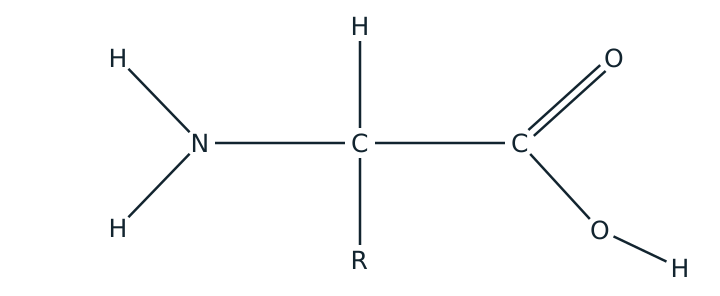
Only the R group differs between one amino acid and another. Everything proteins can do comes from that one variable part: some R groups are charged, some are polar, some are hydrophobic, and some can form bonds with each other. Twenty options, repeated along a chain of hundreds, gives an effectively unlimited number of possible proteins.
The peptide bond
Two amino acids join by a condensation reaction. The carboxyl group of one reacts with the amino group of the other, one molecule of water is released, and a peptide bond forms. Two amino acids joined this way make a dipeptide; many make a polypeptide.
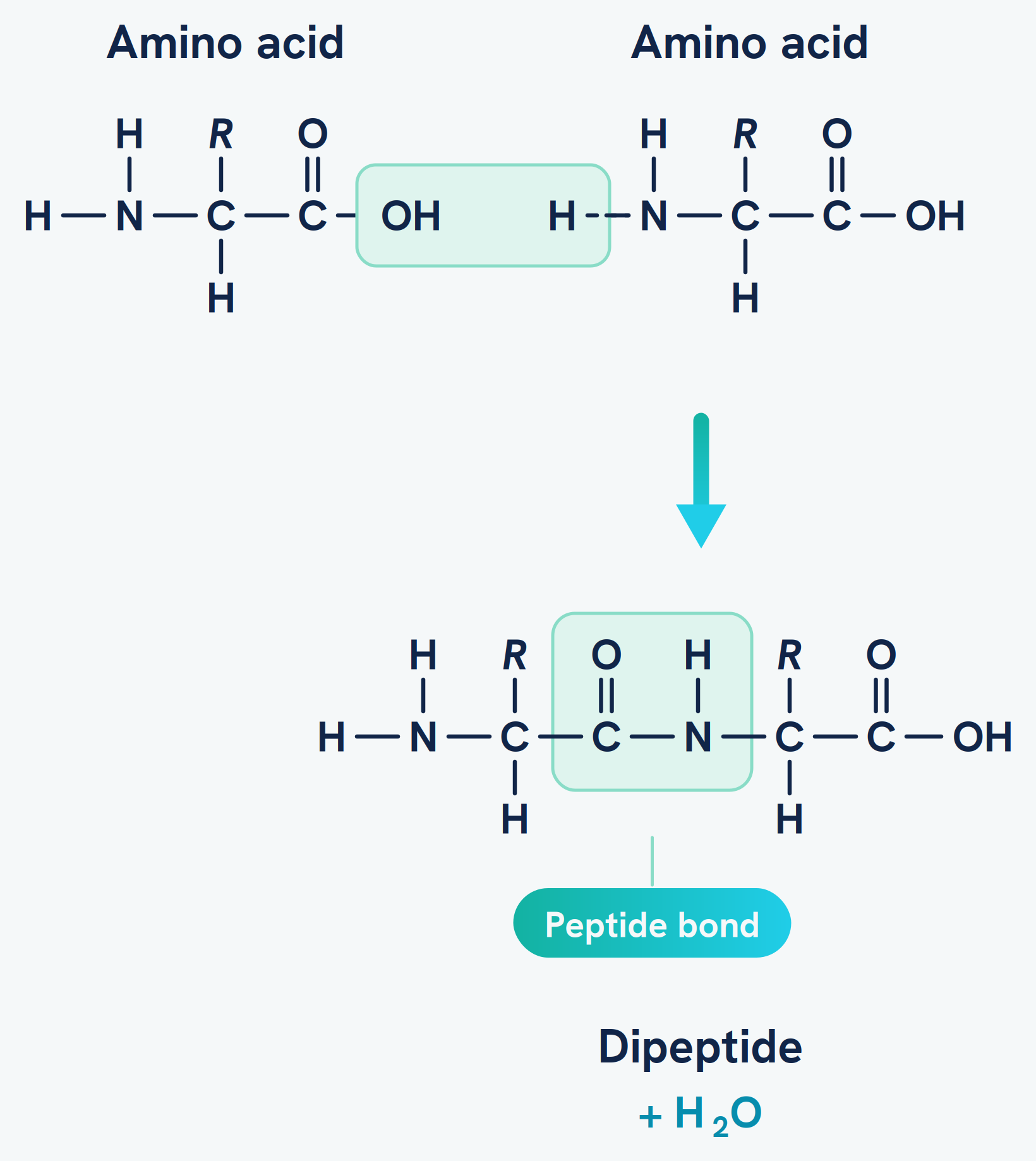
Because the reaction always uses the amino group of one and the carboxyl group of the next, a polypeptide has an amino group free at one end and a carboxyl group free at the other. The chain therefore has a direction, and the order of amino acids along it has to be read one way.
The four levels of protein structure
A protein's shape is built up in stages, and each stage is held together by different bonds. Knowing which bond belongs to which level is the single most examined thing here.
| Level | What it is | Held together by |
|---|---|---|
| Primary | The sequence of amino acids in the chain | Peptide bonds |
| Secondary | Coiling into an alpha helix or folding into a beta pleated sheet | Hydrogen bonds |
| Tertiary | The overall three-dimensional folding of one chain | Hydrogen bonds, ionic bonds and disulfide bridges |
| Quaternary | Two or more polypeptide chains joined together | The same bonds, between chains |
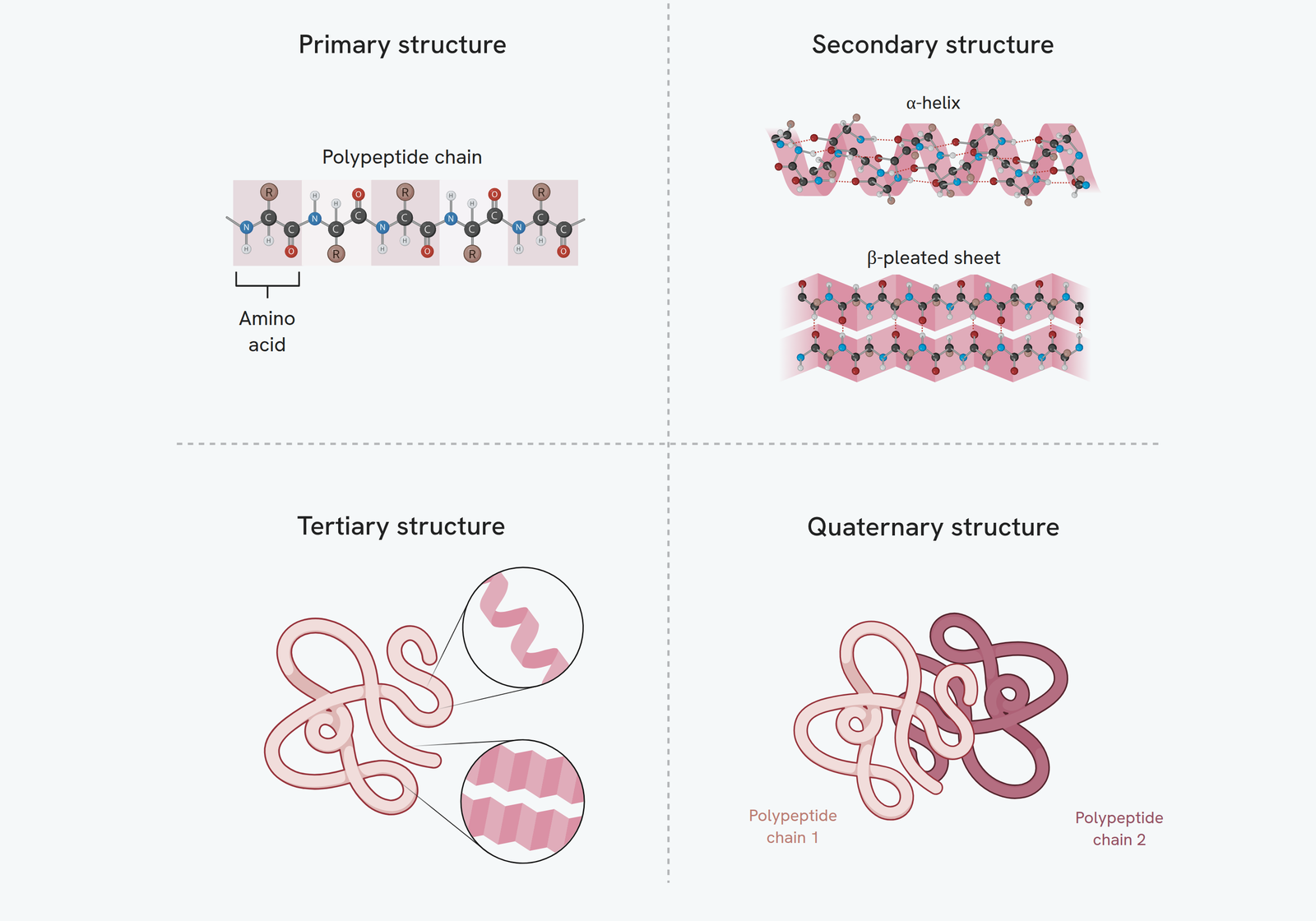
The three bonds at tertiary level are not interchangeable, and questions often ask which is which:
- Hydrogen bonds form between polar R groups. They are individually weak, but there are many of them.
- Ionic bonds form between oppositely charged R groups, and they are broken by a change in pH, because pH changes the charges.
- Disulfide bridges are covalent bonds between two R groups containing sulfur. They are the strongest of the three and the hardest to break.
Not every protein has a quaternary structure. Haemoglobin does, being four polypeptide chains together; a single-chain enzyme does not.
Why the primary sequence decides everything
The R groups are what form the bonds at secondary and tertiary level. Which R groups are present, and where they sit along the chain, is the primary structure. So the primary structure determines which bonds can form and where, and therefore determines the final three-dimensional shape.
That is why changing a single amino acid can matter so much. A different R group in one position can remove a bond that was holding a fold in place, the protein folds differently, and a protein with the wrong shape usually cannot do its job. This is the link to enzymes, where the shape in question is the active site, and to the genetic code, where a change in one base changes one amino acid.
Testing for proteins
Proteins are found with the Biuret test, which detects peptide bonds:
- Add sodium hydroxide solution to make the sample alkaline.
- Add a few drops of dilute copper(II) sulfate solution.
- The pale blue reagent turns purple if protein is present. It stays blue if not.
Since the test detects the peptide bond itself, it finds any polypeptide, whatever its shape. No heating is needed, which sets it apart from the Benedict's test.
How this fits together
Proteins are the group whose shape matters most, and the proteins where it matters most of all are the ones that catalyse reactions. That is the subject of the enzymes guide, which is the rest of this section of the specification.