AQA GCSE Biology: Enzymes and Digestion
A clear GCSE Biology revision guide to enzymes and digestion: the lock and key model, the digestive enzymes, bile, the effect of temperature and pH, and food tests.
Digestion breaks large food molecules into small ones the body can absorb, and enzymes make it happen. This guide explains what enzymes are and how the lock and key model works, the three main digestive enzymes and what they do, the role of bile, how temperature and pH affect enzymes, and the tests used to identify food molecules. The lock and key idea runs through the whole guide.
Digestion and enzymes
Digestion is the breaking down of large, insoluble food molecules into smaller, soluble ones that can be absorbed into the bloodstream.
An enzyme is a biological catalyst: a protein that speeds up a chemical reaction without being used up or permanently changed, so it can be used again and again. Each enzyme catalyses one specific reaction.
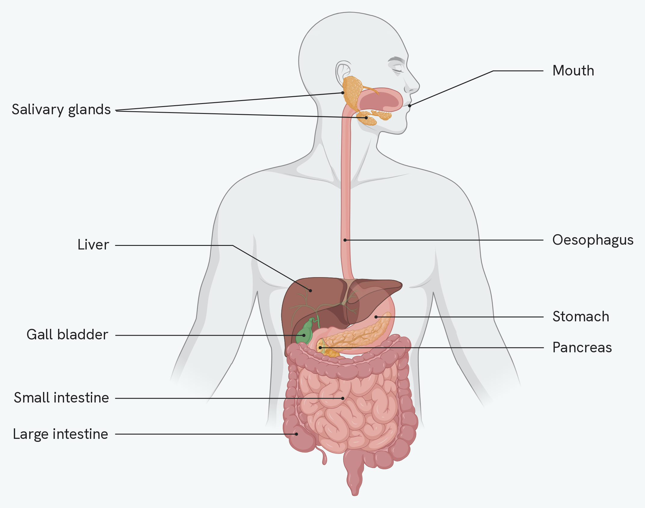
Two kinds of organ are labelled here, and the difference is worth fixing early. The mouth, oesophagus, stomach, small intestine and large intestine form the tube the food actually travels along. The salivary glands, liver, gall bladder and pancreas are not on that path at all: they sit beside it and release their secretions into it.
That distinction explains the table further down. It names where each enzyme is produced and where it acts, and for most of them those are different places.
The lock and key model
An enzyme works because of the shape of its active site.
- The substrate (the molecule the enzyme acts on) fits into the active site, because their shapes are complementary.
- Once bound, the reaction happens, for example the substrate is broken into smaller product molecules.
- The products are released, and the enzyme is unchanged, ready to catalyse another reaction.
Enzymes are specific because each active site has a unique three-dimensional shape, and only a substrate with a complementary shape can fit. Other molecules cannot bind, so the enzyme cannot catalyse their reactions.
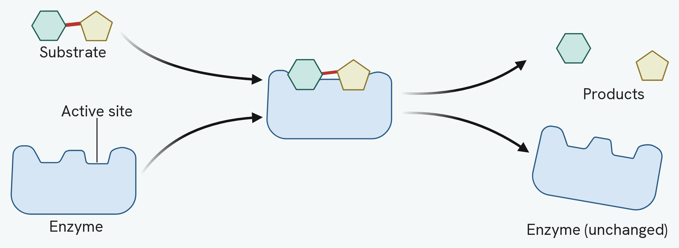
Read the right-hand side of that figure carefully, because it carries two things that are easy to miss. The substrate has been broken into two products, and the enzyme's active site is empty and exactly the shape it was before. The enzyme has not been used up, so it can bind another substrate straight away.
The digestive enzymes
Three groups of enzyme break down the three main food groups.
| Enzyme | Breaks down | Into | Produced in | Acts in |
|---|---|---|---|---|
| Carbohydrase (e.g. amylase) | Carbohydrates (e.g. starch) | Simple sugars (e.g. glucose) | Salivary glands, pancreas, small intestine | Mouth and small intestine |
| Protease | Proteins | Amino acids | Stomach, pancreas, small intestine | Stomach and small intestine |
| Lipase | Lipids (fats) | Fatty acids and glycerol | Pancreas, small intestine | Small intestine |
The products are then used by the body: glucose for respiration and to make new carbohydrates; amino acids to build new proteins for growth and repair; and fatty acids and glycerol to build lipids for cell membranes and energy storage.
Bile
Bile is made by the liver, stored in the gall bladder, and released into the small intestine. It has two jobs.
- It is alkaline, so it neutralises the hydrochloric acid arriving from the stomach, creating the optimum pH for the enzymes in the small intestine.
- It emulsifies lipids, breaking large fat droplets into smaller ones. This increases the surface area for lipase to work on, so fat is digested faster.
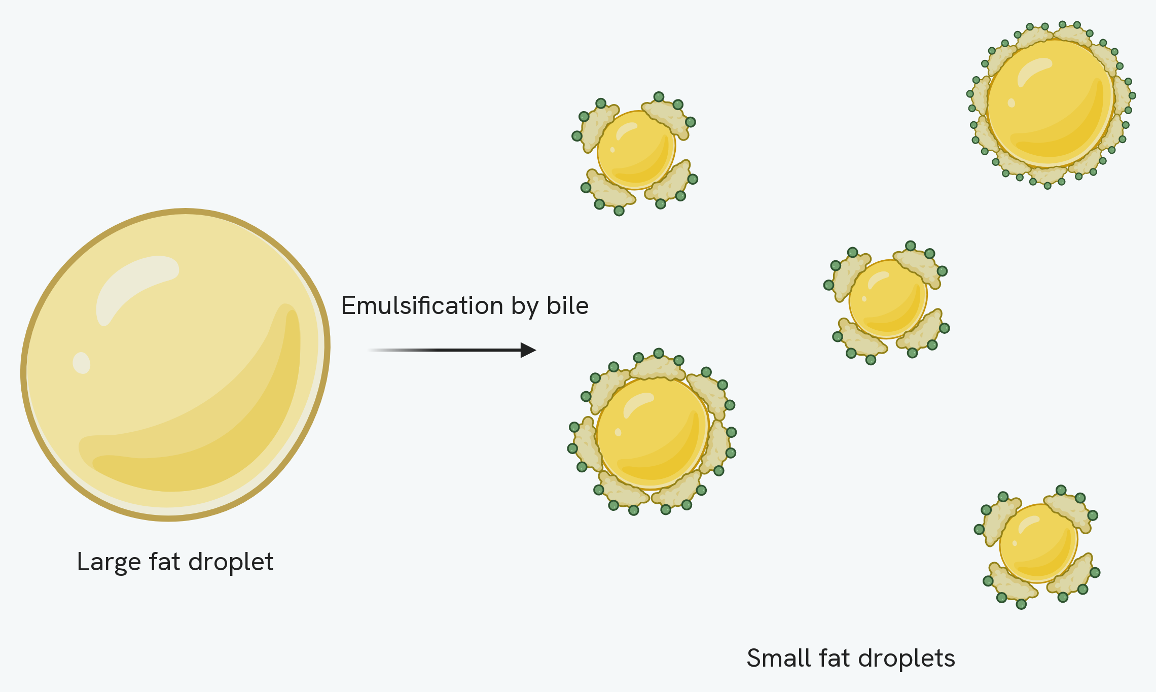
Notice what has not changed. There is the same amount of fat on both sides of that arrow: bile is not an enzyme and it digests nothing. What changes is that the fat is now divided into many small droplets, and many small droplets have a far greater total surface area than one large one. More surface means more places for lipase to work at once, so the fat is digested faster.
Saying bile breaks down fat is the commonest way to lose this mark. Bile breaks fat up; lipase breaks it down.
The effect of temperature and pH
If an enzyme gets too hot, or the pH moves too far from its optimum, its active site changes shape so the substrate can no longer fit. The enzyme can no longer catalyse the reaction; this is denaturation.
- Raising the temperature towards the optimum speeds the reaction up, because molecules have more kinetic energy and collide more often. Above the optimum, the rate falls sharply because the enzyme denatures.
- Moving the pH above or below the optimum slows the reaction, because the enzyme denatures. Different enzymes have different optimum pH values: stomach protease works best at about pH 2, while pancreatic enzymes work best at about pH 7 to 8.
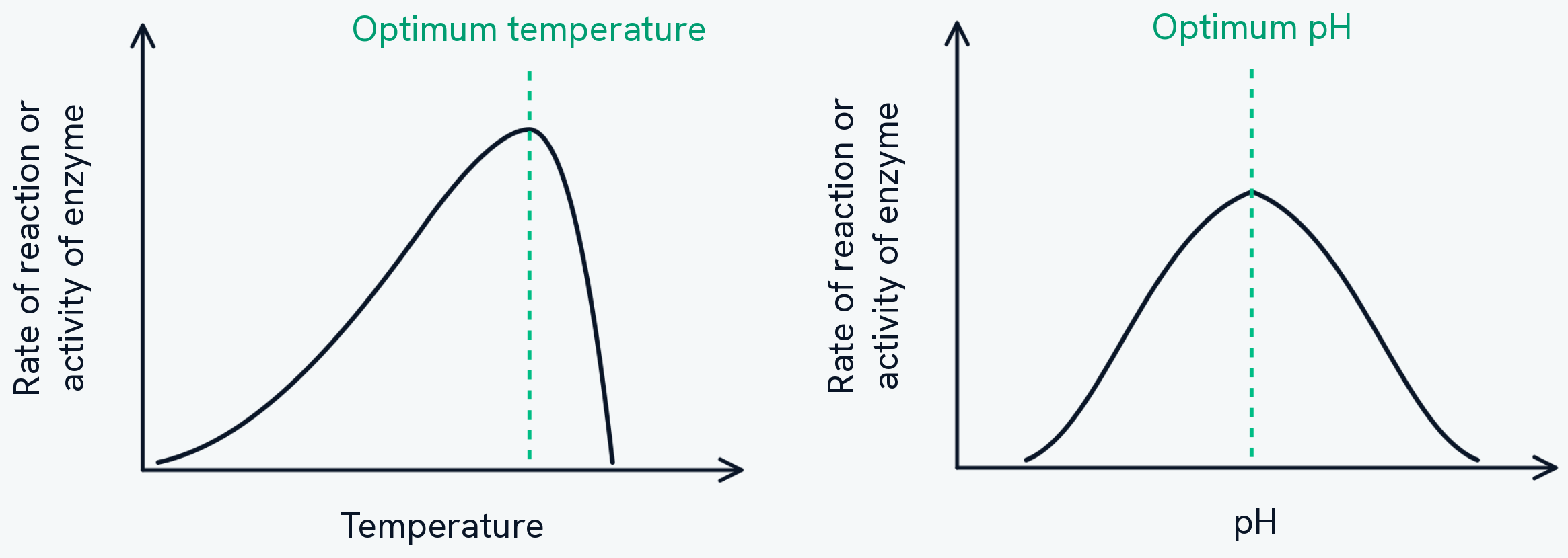
The two curves are not the same shape, and the difference carries a mark. The temperature curve rises gradually and then drops very steeply, because warming an enzyme speeds it up right up to the point where it denatures, and a denatured enzyme does not recover. The pH curve is roughly symmetrical, because moving away from the optimum in either direction distorts the active site in much the same way.
Notice too that neither axis has numbers on it. That is deliberate: the optimum is not one fixed value. Stomach protease peaks at about pH 2 and pancreatic enzymes at about pH 7 to 8, so a graph shaped like this describes enzymes in general rather than any particular one.
Testing for food molecules
Simple tests show which food molecules are present, each giving a colour change.
- Sugars (reducing): add Benedict's solution and heat. A positive result changes from blue through green, yellow and orange to brick red, with a redder colour meaning more sugar.
- Starch: add iodine solution. A positive result turns from orange-brown to blue-black.
- Proteins: add Biuret reagent. A positive result changes from blue to purple (lilac).
- Lipids: grind the sample with ethanol, then add water. A positive result forms a milky-white emulsion.
How this fits together
Once food is digested and absorbed, the small molecules are carried around the body by the blood. The pump that moves that blood is the subject of the heart and blood vessels guide.