AQA GCSE Biology: Plant Tissues
A clear GCSE Biology revision guide to plant tissues: the structure of a leaf and the job of each tissue, and the detail of xylem and phloem.
A leaf is an organ made of several tissues, each adapted to help the plant photosynthesise. This guide works through the tissues in a leaf and what each does, then looks in more detail at the transport tissues, xylem and phloem. The theme is that each tissue's structure suits its function.
The tissues in a leaf
A leaf is made up of several tissues, arranged so that light reaches the photosynthetic cells and gases can move in and out.
- Upper and lower epidermis: a single layer of transparent cells that lets light through to the cells beneath. The upper epidermis makes a waxy cuticle that waterproofs the leaf and reduces water loss.
- Palisade mesophyll: near the upper surface, these cells are packed with chloroplasts to absorb as much light as possible for photosynthesis.
- Spongy mesophyll: below the palisade layer, this tissue has many air spaces that give a large internal surface area for the rapid diffusion of gases (carbon dioxide in, oxygen out).
- Guard cells and stomata: guard cells are pairs of cells, mainly on the lower epidermis, that control the opening and closing of stomata, the small pores that let gases in and out and through which water vapour is lost.
- Xylem and phloem: the transport tissues, running through the veins, which carry water and minerals (xylem) and dissolved sugars (phloem).
- Meristem tissue: at the root and shoot tips, made of undifferentiated stem cells that can divide and differentiate into any plant cell, allowing continued growth.
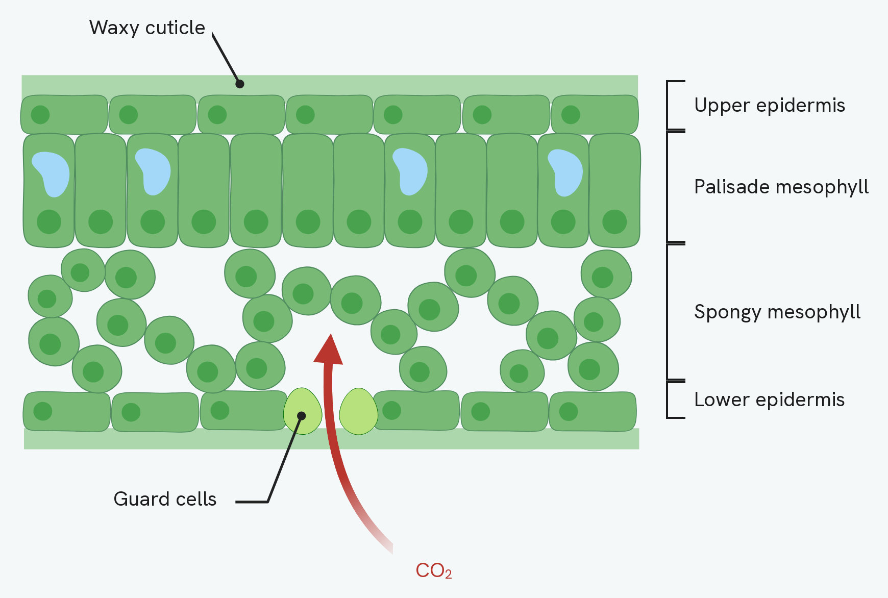
Read that section from the top down and it follows the list above. The waxy cuticle is the clear strip over the surface. Under it the upper epidermis is one layer of cells with no chloroplasts, so light passes straight through to the palisade mesophyll, where the cells stand on end, packed close together and full of chloroplasts. Below that the spongy mesophyll is loosely packed, and the gaps between those cells are the air spaces that let gases move about inside the leaf.
The two pale cells in the lower epidermis are the guard cells, and the gap between them is a stoma. The red arrow is carbon dioxide diffusing in through it, which is what all those air spaces above are for.
One thing this section does not show is a vein. The cut has been made between two veins, so there is no xylem or phloem anywhere in it. Those are the subject of the next section.
Xylem and phloem in detail
(Triple award only.) The two transport tissues are built very differently, to suit what they carry.
Xylem transports water and dissolved mineral ions from the roots to the leaves. Its cells are strengthened with lignin to withstand the pressure of moving water, they are hollow with no cytoplasm so water flows freely, and their end walls have broken down to form long continuous tubes. Xylem cells are dead.
Phloem transports dissolved sugars (such as sucrose) from the leaves to the rest of the plant, both upwards and downwards. Its cells are living, have no nucleus and few other structures to make room for the sugars, and their end walls have pores (sieve plates) that let sugars pass from cell to cell.
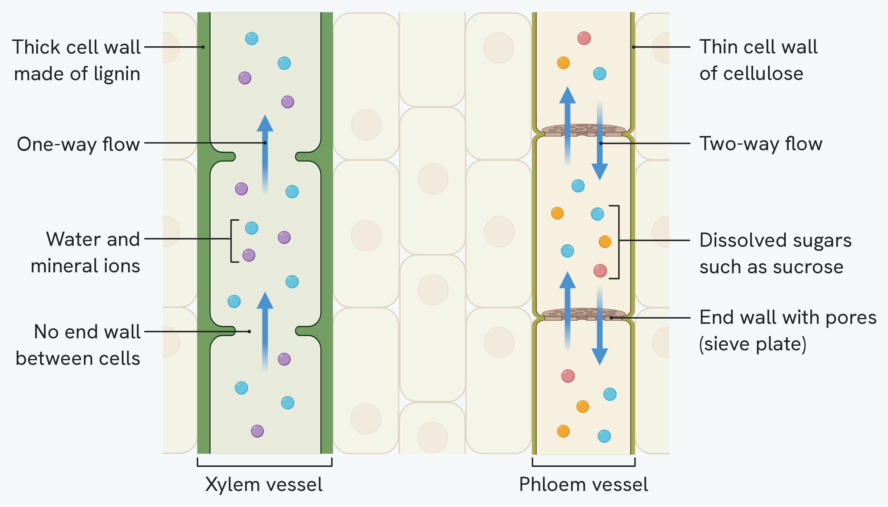
Side by side the differences are easier to hold on to. The xylem on the left is a hollow tube: thick walls stiffened with lignin, no end walls at all between its cells, and water and mineral ions moving in one direction only, up from the roots. The phloem on the right is alive: thin cellulose walls, and end walls that are still there but pierced with pores, which is the sieve plate. Sugars travel through it in both directions.
The strip of plain cells between the two is not something you are asked to name. It is there because xylem and phloem sit alongside each other in the same vein.
How this fits together
These tissues, especially xylem and phloem, are the plant's transport system. How water and sugars actually move through them, in transpiration and translocation, is the subject of the plant transport guide.