AQA GCSE Biology: The Heart and Blood Vessels
A clear GCSE Biology revision guide to the heart and blood vessels: the double circulatory system, the path of blood through the heart, the pacemaker, and arteries, veins and capillaries.
The heart pumps blood around the body, carrying oxygen and food to every cell. This guide explains the double circulatory system, the journey of blood through the heart, how the heartbeat is controlled, and how the three types of blood vessel are each built for their job. The link to hold onto is that structure matches function throughout.
The double circulatory system
The heart is a muscular organ that pumps blood around a double circulatory system: blood passes through the heart twice for each complete circuit of the body.
- The right side pumps deoxygenated blood to the lungs, where it gains oxygen and loses carbon dioxide.
- The left side pumps the returning oxygenated blood to the rest of the body.
The path of blood through the heart
Following the blood through in order:
- Deoxygenated blood from the body enters the right atrium through the vena cava.
- It flows into the right ventricle, which pumps it to the lungs through the pulmonary artery.
- In the lungs the blood is oxygenated.
- It returns to the left atrium through the pulmonary vein.
- It flows into the left ventricle, which pumps it to the body through the aorta.
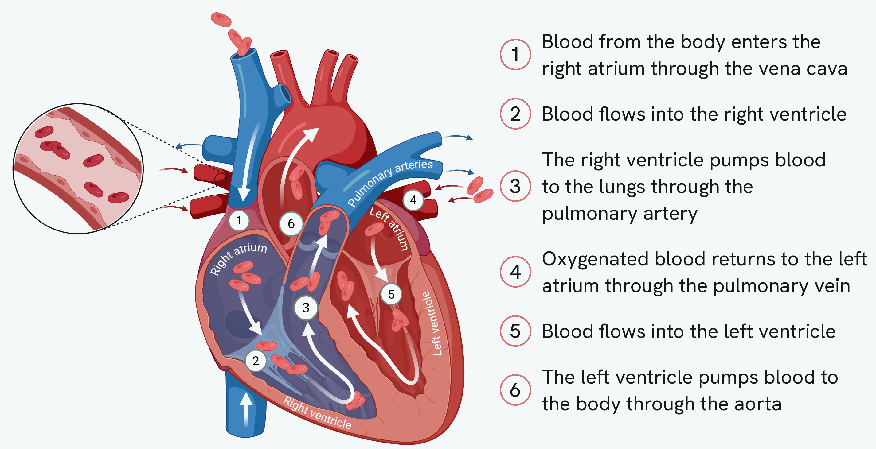
The numbered steps beside the drawing are the same route as the list above, so read one against the other until the order sticks.
Two things on that drawing are worth stopping at. The first is which side is which: the heart's right side is drawn on your left, because a heart is always labelled from the point of view of the person it belongs to, not the person looking at it. Almost every mistake in this topic starts there.
The second is that the two sides never mix. The deoxygenated blood on the right and the oxygenated blood on the left are kept apart by a wall of muscle down the middle, which is what makes the double circulatory system possible: blood cannot take a short cut back to the body without going to the lungs first.
The left ventricle wall is thicker than the right, because it has to pump blood all the way around the body, which needs a much greater force than pumping the short distance to the lungs.
The pacemaker
A group of cells in the right atrium acts as the heart's natural pacemaker. It sends out regular electrical impulses that make the heart muscle contract rhythmically. If the natural pacemaker becomes faulty, an artificial pacemaker, an electrical device fitted in the chest, can be used to keep the heartbeat regular.
The blood vessels
There are three types of blood vessel, each adapted to its role.
| Vessel | Job | Key features |
|---|---|---|
| Artery | Carries blood away from the heart at high pressure | Thick walls with muscle and elastic tissue; narrow lumen |
| Vein | Carries blood back to the heart at low pressure | Thinner walls; large lumen; valves to stop backflow |
| Capillary | Allows exchange of substances with cells | Walls one cell thick (short diffusion distance); narrow lumen |
Arteries have thick, muscular and elastic walls to withstand and maintain the high pressure by stretching and recoiling, and a narrow lumen to help keep pressure high. Veins carry blood at low pressure, so they have thinner walls and a large lumen to reduce resistance, plus valves to keep blood moving towards the heart. Capillaries have walls just one cell thick for a short diffusion distance, and a narrow lumen that slows the blood so there is more time for substances such as oxygen and glucose to diffuse into the cells.
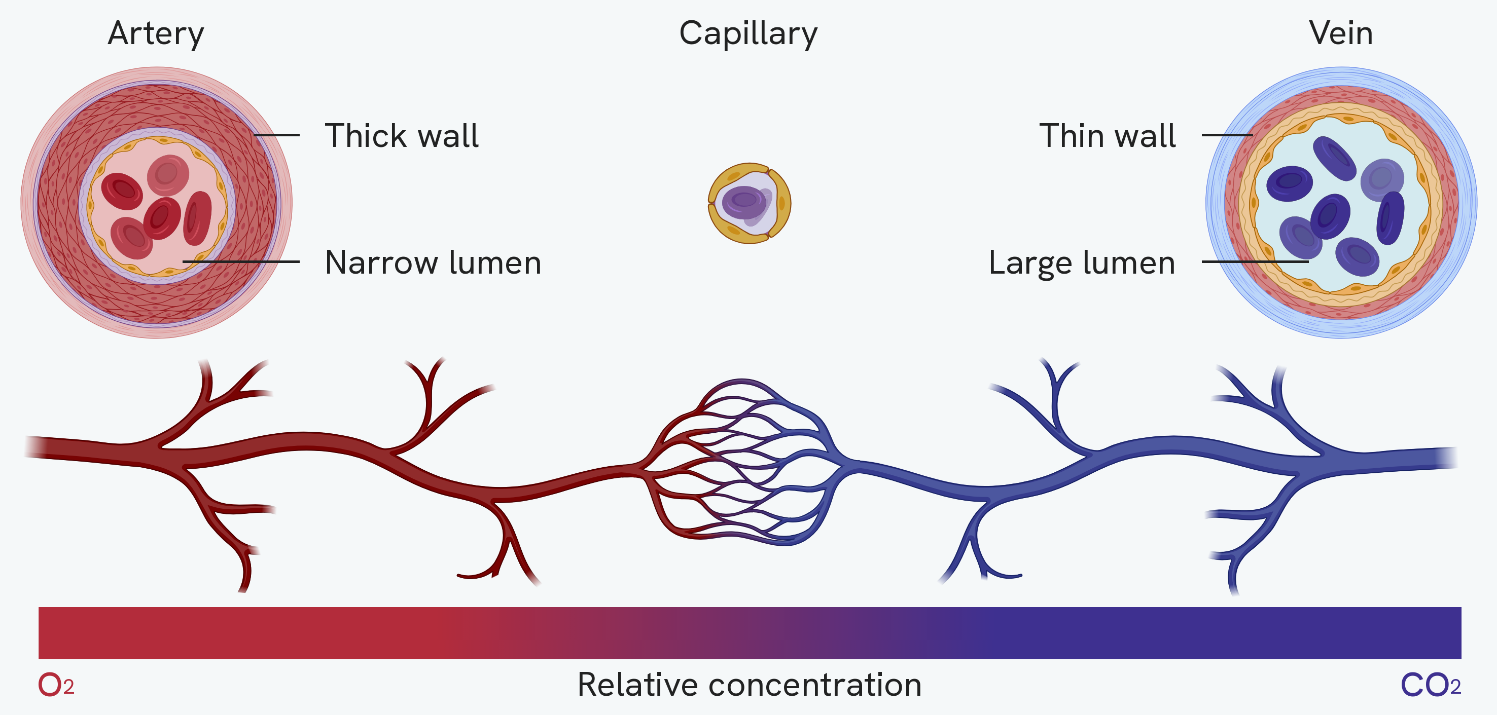
Cut across and set side by side, the table becomes something you can see. The artery is nearly all wall around a small lumen; the vein is nearly all lumen inside a thin one. All three are drawn at the same scale, which is why the capillary looks so small next to them. It really is that small, a ring one cell thick, and that single-cell wall is what makes it the vessel where exchange happens.
The vessel running beneath them shows something the table cannot, which is the order. Blood leaves the heart in an artery, the artery divides until it becomes a network of capillaries, and those join back up into a vein that carries it to the heart. Every artery ends in a capillary network, and every vein begins at one.
Follow the colour across, and the bar with it. The blood arrives red and leaves blue, because the capillaries are where oxygen diffuses out into the cells and carbon dioxide diffuses in. That is why the exchange row of the table belongs to the capillary and not to the other two: only there is the wall thin enough, and the blood slow enough, for it to happen.
How this fits together
The vessels carry the blood, but the blood itself is a tissue with several components, each with its own job, which is the subject of the blood guide.