AQA A-Level Biology: Gene Expression and Cancer
A clear revision guide to cancer for AQA A-Level Biology: benign and malignant tumours, tumour suppressor genes and oncogenes, epigenetics in cancer, and oestrogen in breast cancer.
Cancer is a disease of gene expression: it happens when the genes that control cell division are damaged or wrongly regulated, so cells divide out of control. This guide explains how tumours form, the difference between benign and malignant ones, the two kinds of gene that normally control division, and how both genetic mutations and epigenetic changes can disrupt them. It ends with the role of oestrogen in some breast cancers. The unifying idea is loss of control over the cell cycle.
How tumours form
Cell division is normally tightly controlled. Mutations in the genes that control mitosis can cause uncontrolled cell division, and if this produces a mass of abnormal cells, a tumour forms.
Tumours are either benign or malignant, and the distinction matters clinically.
| Feature | Benign | Malignant (cancerous) |
|---|---|---|
| Growth | Usually slow | Usually faster |
| Differentiation | Cells well differentiated | Cells poorly differentiated |
| Nuclei | Normal, regular | Irregular, larger or darker |
| Borders | Well defined, often within a capsule, so do not invade | Poorly defined, no capsule, so invade surrounding tissue |
| Metastasis | Does not spread | Spreads by metastasis, forming secondary tumours |
| Treatment | Usually removed by surgery, rarely returns | Surgery plus radiotherapy or chemotherapy, often returns |
Metastasis is when cells break off a malignant tumour, travel to other parts of the body, and form secondary tumours there.
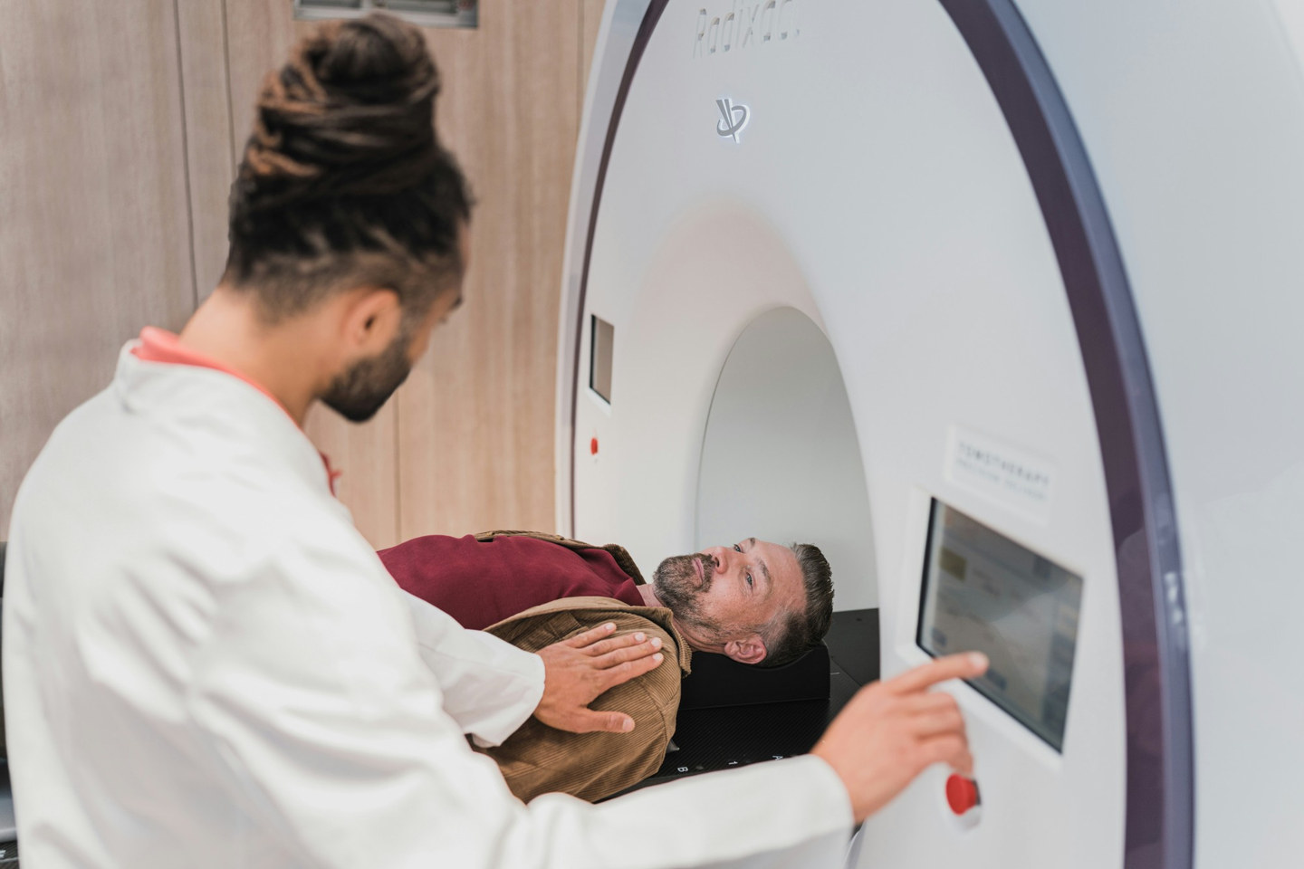
Radiotherapy is the treatment named in the last row of that table, and it works on the same fact the rest of this guide turns on. Ionising radiation does most damage to cells that are dividing, and the cells of a tumour divide far more often than the cells around them, so the tumour is hit hardest.
Tumour suppressor genes and oncogenes
Two kinds of gene normally control cell division, and cancer results when they go wrong.
Tumour suppressor genes code for proteins that slow the cell cycle. They inhibit cell division when DNA damage is detected, or cause the self-destruction of a potential tumour cell if the damage cannot be repaired. An example is p53. They can be lost in two ways:
- a mutation produces a non-functional protein, because the changed amino acid sequence alters the protein's tertiary structure; or
- an epigenetic change, increased DNA methylation or decreased histone acetylation, stops the gene being transcribed, so the protein is not made.
Either way, the brake on cell division is lost and division becomes uncontrolled.
Proto-oncogenes code for proteins that stimulate cell division. An oncogene is a mutated or abnormally expressed version of a proto-oncogene, and it drives division too hard. This happens when:
- a mutation causes the protein to be overproduced or permanently activated, again through a change in tertiary structure; or
- an epigenetic change, decreased DNA methylation or increased histone acetylation, increases transcription and so overproduces the protein.
Either way, the accelerator on cell division is jammed on.
A key point is that a tumour needs both alleles of a tumour suppressor gene to be faulty, but only one allele of a proto-oncogene to become an oncogene. This is because a single working tumour suppressor allele can still make enough protein to control the cell cycle, whereas a single oncogene allele already makes enough protein to drive uncontrolled division. (Note that all cells have tumour suppressor genes; cancer comes from the loss of the tumour suppressor protein, not the gene.)
Epigenetics and cancer treatment
Because some cancers are driven by epigenetic changes rather than mutations, and epigenetic changes can in principle be reversed, they offer a route to treatment. A drug could:
- increase DNA methylation or decrease histone acetylation of an oncogene, to inhibit its transcription; or
- decrease DNA methylation or increase histone acetylation of a tumour suppressor gene, to restore its transcription.
Either change would help bring cell division back under control.
Oestrogen and breast cancer
Some breast cancers are oestrogen receptor-positive, meaning increased oestrogen can drive them.
- These cancer cells have oestrogen receptors, which are inactive transcription factors.
- When oestrogen concentration rises, more oestrogen binds, forming more oestrogen-receptor complexes, which are active transcription factors.
- These bind to the promoter regions of genes that code for proteins stimulating cell division.
- This increases expression of those genes, so the rate of cell division increases.
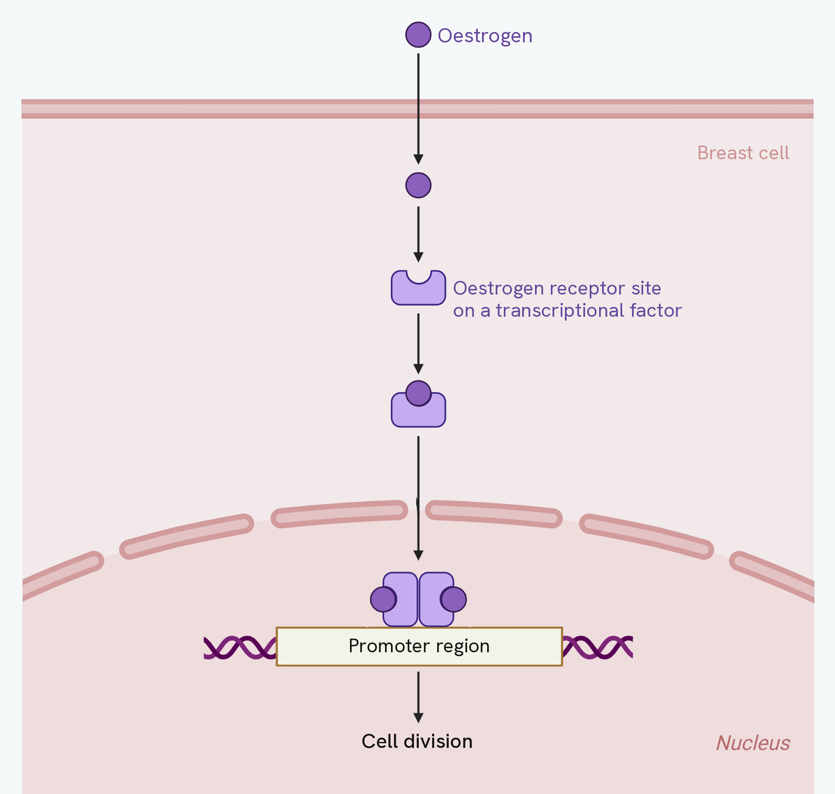
Four steps on that diagram, and each one answers a different question an examiner can ask.
Oestrogen gets in without any help, because it is a lipid-soluble steroid and the membrane is made of lipid. Nothing carries it and nothing lets it through; it simply dissolves across. That is why the first arrow passes straight through the membrane rather than stopping at a channel.
What it binds to in the cytoplasm is not a receptor on its own. It is a receptor site on a transcriptional factor, which is why binding matters: the factor changes shape and its DNA-binding site becomes active. Oestrogen never touches the DNA itself.
The complex then binds the promoter region of the gene. Not the gene, the promoter, which is the stretch in front of it where transcription is started.
And the arrow at the bottom is the whole reason this sits in a guide about cancer. The genes being switched on are ones that stimulate cell division. More oestrogen means more active transcriptional factors, which means more of those genes transcribed, which means faster division. That is the chain a question about increased oestrogen exposure is asking you to follow.
This suggests a treatment: drugs with a similar structure to oestrogen can bind to the oestrogen receptors and block oestrogen from binding. With fewer active transcription factors formed, fewer of the genes driving the cell cycle are switched on.
How this fits together
Cancer shows what happens when control of gene expression fails at the level of cell division, and it also shows how understanding that control points the way to treatment. Studying genes and their expression on this scale has been transformed by reading whole genomes, which is the subject of the genome projects guide.