AQA A-Level Biology: Digestion and Absorption
A clear revision guide to digestion and absorption for AQA A-Level Biology: how carbohydrates, lipids and proteins are hydrolysed, and how the products are absorbed in the ileum.
The food we eat is made of large molecules that are far too big to be absorbed. Digestion solves this by hydrolysing them into small, soluble molecules that can cross cell membranes into the blood. This guide covers how each food group is digested and how the products are absorbed.
What digestion is
Digestion is the hydrolysis of large, insoluble biological molecules into smaller, soluble ones that are small enough to be absorbed across cell membranes into the blood. Each type of molecule is broken by a specific enzyme that adds water to split a particular bond.
Digesting carbohydrates
Starch is digested in two stages. First, amylase, made by the salivary glands and the pancreas, hydrolyses starch into maltose. Then a membrane-bound enzyme, maltase, attached to the cells lining the ileum, hydrolyses maltose into glucose. Both steps break a glycosidic bond.
Other disaccharides are dealt with by their own membrane-bound disaccharidases:
- maltase hydrolyses maltose into glucose and glucose
- sucrase hydrolyses sucrose into glucose and fructose
- lactase hydrolyses lactose into glucose and galactose
Digesting lipids
Lipids are not soluble in water, which makes them hard for enzymes to reach, so they are first emulsified. Bile salts, made by the liver, break large lipid droplets into many smaller droplets. This increases the surface area of the lipids, so the enzyme can work faster. It is worth being clear that bile salts are not enzymes: they emulsify lipids but do not digest them.
Lipase, made in the pancreas, then hydrolyses the lipids (such as triglycerides) into monoglycerides and fatty acids, breaking the ester bonds.
Digesting proteins
Proteins are digested by a team of enzymes working together. Endopeptidases hydrolyse peptide bonds within the polypeptide chain, cutting it into shorter peptides. This creates many more ends, and therefore more surface area, for the next enzymes to work on. Exopeptidases then hydrolyse the peptide bonds at the ends of these peptides, removing amino acids one at a time. Finally, membrane-bound dipeptidases split the bond in any remaining dipeptides into single amino acids. Every step breaks a peptide bond.
Why membrane-bound enzymes help
Maltase, the disaccharidases and dipeptidases are all membrane-bound, fixed to the cell-surface membranes of the epithelial cells lining the ileum. This matters because they release the final products, glucose and amino acids, right at the site of absorption. This keeps the concentration of these products high next to the cells, maintaining the gradient that drives absorption.
Absorbing amino acids and monosaccharides
Glucose, other monosaccharides and amino acids are absorbed from the ileum by co-transport with sodium ions. It works in three steps:
- Sodium ions are actively transported out of the epithelial cell into the blood, using ATP. This keeps the sodium ion concentration inside the cell low, lower than in the gut lumen.
- Sodium ions then diffuse from the lumen into the epithelial cell down this gradient, through a co-transporter protein that carries a glucose molecule or amino acid in at the same time, even though that molecule is moving against its own concentration gradient.
- The glucose or amino acid, now concentrated inside the cell, moves into the blood by facilitated diffusion.
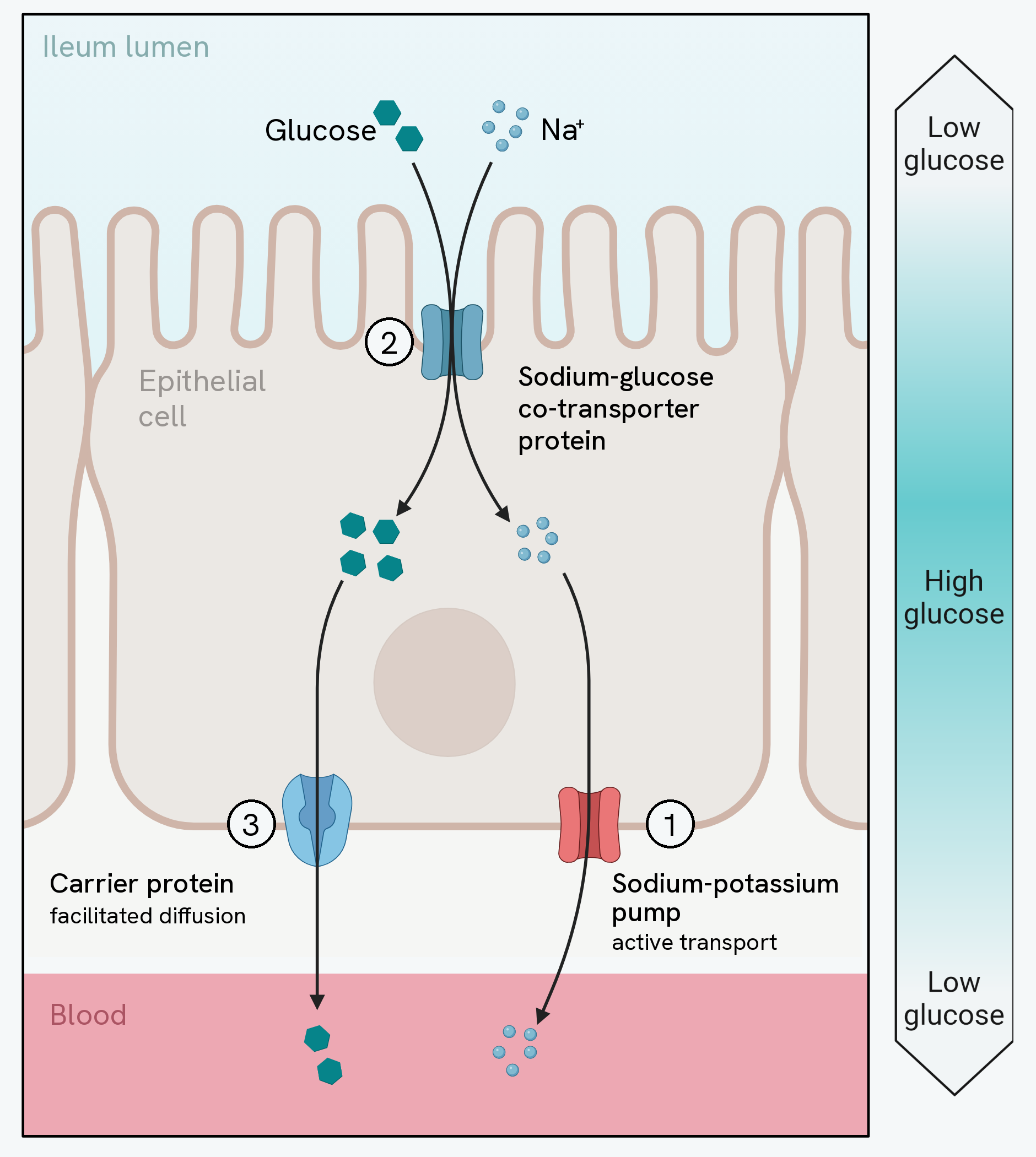
The numbers on that diagram are the three steps above, and they run bottom, top, bottom rather than down the page. Three different proteins are involved, and knowing which membrane each one sits in is most of the answer.
The sodium-potassium pump is in the membrane at the base of the cell, facing the blood. It is the only one of the three that uses ATP, and everything else depends on it: by pumping sodium out, it keeps the sodium concentration inside the cell low.
The co-transporter protein is in the membrane at the top of the cell, facing the lumen. Sodium diffuses through it down the gradient the pump has created, and glucose is carried through at the same time. This is the step where glucose moves against its own concentration gradient, and notice that it uses no ATP of its own.
The carrier protein is back at the base, and glucose leaves through it by facilitated diffusion, down its gradient into the blood.
The bar beside the cell is the reason any of this is needed. Glucose is low in the lumen, high inside the cell, and low in the blood again. Diffusion on its own could never move it from the first of those to the second.
This is why anything that stops the sodium pump, for example a poison that blocks active transport, also stops glucose absorption: without the sodium gradient, the co-transporter cannot pull glucose into the cell.
Absorbing lipids: the role of micelles
The products of lipid digestion are absorbed differently. Bile salts combine with the monoglycerides and fatty acids to form tiny structures called micelles. Micelles make the products more soluble in water and carry them to the epithelial cells lining the ileum, keeping a high concentration of fatty acids and monoglycerides right next to the cells.
At the cell, the micelle breaks up and releases its contents. Because monoglycerides and fatty acids are lipid-soluble, they diffuse straight across the cell membrane into the epithelial cell. It is the products, not the whole micelle, that are absorbed.
Inside the cell the triglycerides are reformed, then packaged with proteins into structures called chylomicrons. These are moved to the cell membrane in vesicles and released by exocytosis, entering the lymphatic system before eventually joining the blood.
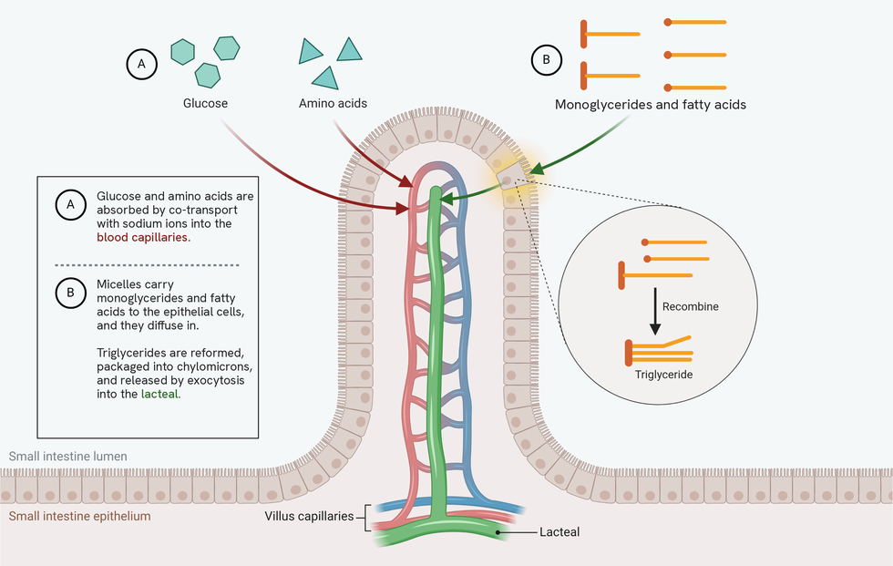
Both routes are on that one villus, and the point of putting them together is that they end in different vessels.
Glucose and amino acids go into the blood capillary, which carries them away in the blood. The products of lipid digestion go into the lacteal, which is part of the lymphatic system, not the blood; they only reach the bloodstream later, when the lymph drains into it. If a question asks where an absorbed product goes, the answer depends entirely on which of the two it is.
The magnified circle is worth reading carefully, because it is drawn the way the specification wants it. What recombines is a monoglyceride and two fatty acids, not glycerol and three fatty acids. Glycerol and fatty acids is the GCSE answer; the products AQA names at A-Level are monoglycerides and fatty acids, and that is what a mark scheme will be looking for here.