AQA A-Level Biology: The Heart and Circulation
A clear revision guide to the heart and circulation for AQA A-Level Biology: the double circulatory system, the cardiac cycle and valves, blood vessels, and tissue fluid formation.
A large animal cannot rely on diffusion to move substances around, so it needs a mass transport system: blood, pumped by a heart, through a network of vessels. This guide covers how mammalian circulation is arranged, how the heart beats, how the blood vessels are built for their jobs, and how tissue fluid forms.
The double circulatory system
Mammals have a closed, double circulatory system. Closed means the blood stays inside vessels; double means the blood passes through the heart twice for every complete circuit of the body. One loop carries deoxygenated blood from the right side of the heart to the lungs, and returns oxygenated blood to the left side. The other loop carries oxygenated blood from the left side to the rest of the body, and returns deoxygenated blood to the right side.
This arrangement has two advantages. It keeps oxygenated and deoxygenated blood separate, so the blood delivered to the body is fully loaded with oxygen. And because blood is pumped again after returning from the lungs at low pressure, it can be sent to the body at high pressure, so substances are delivered and removed quickly.
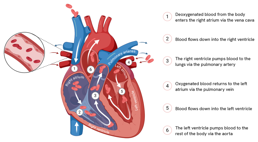
Trace that path once and the double circulation stops being two facts to remember. Steps one to three are the loop to the lungs, steps four to six the loop to the body, and the two never meet because the septum divides the heart down the middle. Each half of the heart is doing the same job as the other, to a different destination.
The main blood vessels
Several named vessels carry blood to and from the heart, lungs and other organs:
| Vessel | Carries | Between |
|---|---|---|
| Vena cava | Deoxygenated blood | Body tissues to the heart |
| Pulmonary artery | Deoxygenated blood | Heart to the lungs |
| Pulmonary vein | Oxygenated blood | Lungs to the heart |
| Aorta | Oxygenated blood | Heart to the body |
| Renal artery | Oxygenated blood | Aorta to the kidneys |
| Renal vein | Deoxygenated blood | Kidneys to the vena cava |
| Coronary arteries | Oxygenated blood | Aorta to the heart muscle itself |
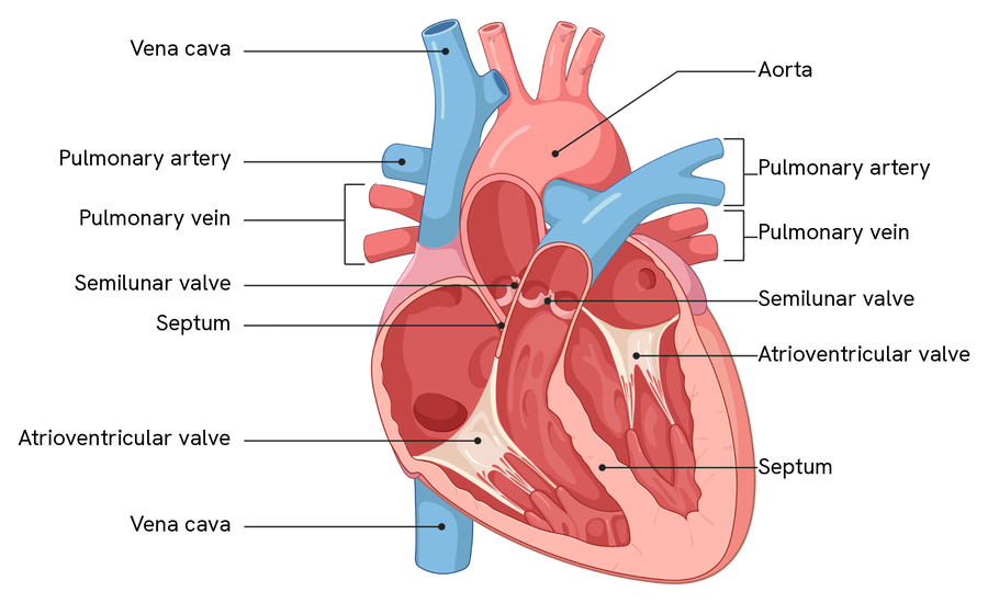
Several names appear twice on that diagram, and that is correct rather than an oversight. There are two atrioventricular valves, one between the atrium and ventricle on each side, and two semilunar valves, one where each artery leaves. The vena cava is labelled where it enters above and again where it enters below.
You will meet other names for the same structures elsewhere. The atrioventricular valves are often called the tricuspid (right) and bicuspid or mitral (left), and the semilunar valves the pulmonary and aortic valves. Those are not wrong, but AQA asks for atrioventricular and semilunar, so write those.
For the four chambers, look back at the blood flow diagram above, where each is named. One point there is worth carrying over, because it catches people out every year: the heart is drawn as it faces you, so the right atrium and ventricle appear on the left of the diagram, and the left ones on the right. Label them from the patient's point of view, not the page's.
The left ventricle has a much thicker muscular wall than the right. This is because it must contract with greater force to generate the high pressure needed to pump blood all the way around the body, while the right ventricle only pumps to the nearby lungs.
The cardiac cycle
The cardiac cycle is one complete heartbeat, and it keeps blood flowing in one direction by opening and closing valves in response to pressure. The rule to hold onto is simple: a valve opens when the pressure behind it is higher than the pressure in front of it, and closes when the pressure in front becomes higher, which stops blood flowing backwards.
There are three stages:
- Atrial systole: the atria contract, so their volume falls and their pressure rises. This pushes the pressure in the atria above that in the ventricles, opening the atrioventricular valves, and blood is forced into the ventricles.
- Ventricular systole: the ventricles contract, raising their pressure above that in the atria, which closes the atrioventricular valves. When ventricular pressure rises above the pressure in the arteries, the semilunar valves open and blood is forced out into the aorta and pulmonary artery.
- Diastole: the atria and ventricles relax, so their volume rises and pressure falls. The semilunar valves close as artery pressure now exceeds ventricular pressure, preventing backflow, and blood flows back into the atria from the veins and passively down into the ventricles.
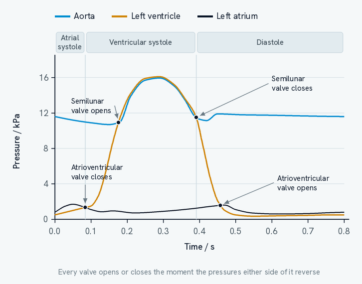
Every one of the four marked points on that graph is a place where two traces cross, and that is the whole trick to reading it. A valve has blood on both sides, and it is pushed open or shut by whichever side has the higher pressure. So wherever two lines swap over, a valve must change state.
Follow the ventricle trace, in amber, around the cycle.
- At 0.08 s it rises past the atrium trace, so blood would be pushed back towards the atrium. The atrioventricular valve closes.
- At 0.18 s it rises past the aorta trace, so blood can now be pushed out into the aorta. The semilunar valve opens, and ejection begins. Notice the ventricle stays slightly above the aorta for the whole of ejection: that difference is what keeps blood moving.
- At 0.39 s the relaxing ventricle falls back below the aorta, so the semilunar valve closes, preventing backflow from the artery.
- At 0.46 s it falls below the atrium, so the atrioventricular valve opens and the ventricle starts filling again.
Between 0.08 s and 0.18 s both valves are shut, so the ventricle is contracting without any blood leaving it. That is why the pressure climbs so steeply there.
To read a cardiac cycle graph, compare the pressures in the two chambers or vessels either side of a valve: the valve is closed whenever the pressure in front of it is higher (preventing backflow), and open when the pressure behind it is higher (letting blood through).
Two useful calculations come from the cycle. Heart rate in beats per minute is 60 divided by the length of one cardiac cycle in seconds. On the graph above one full cycle takes 0.8 s, giving a heart rate of 60 ÷ 0.8 = 75 beats per minute. Cardiac output, the volume of blood pumped per minute, is the stroke volume (the volume pumped per beat) multiplied by the heart rate.
How blood vessels are adapted
Each type of vessel is built for the pressure it handles.
Arteries carry blood away from the heart at high pressure. They have a thick layer of elastic tissue that stretches when the ventricles contract and recoils when they relax, which smooths out the surges and maintains a high pressure. A layer of smooth muscle helps control flow, a thick wall withstands the pressure, and a narrow lumen helps keep the pressure high.
Arterioles are smaller vessels that direct blood into different tissues. They have a thicker layer of smooth muscle than arteries: when it contracts the lumen narrows (vasoconstriction) and reduces blood flow, and when it relaxes the lumen widens (vasodilation) and increases flow.
Capillaries are where exchange happens. Their wall is a single layer of endothelial cells, giving a very short diffusion distance, they form vast branched capillary beds with a huge surface area, and their narrow lumen slows the blood so there is more time for exchange. Small gaps between the cells let fluid and small molecules leave.
Veins return blood to the heart at low pressure. They have a wide lumen to reduce resistance, little elastic or muscle tissue because the pressure is low, and valves to stop blood flowing backwards.
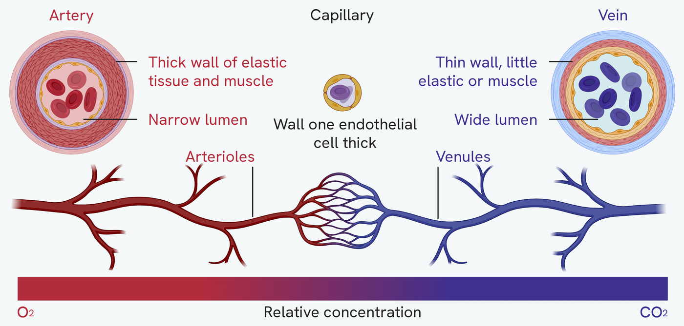
All three cross-sections are drawn to the same scale, and that is the first thing to take from the figure. The capillary is not a smaller drawing of a vessel. It really is that small beside an artery, which is what lets it run between individual cells.
Wall thickness tracks pressure. An artery leaves the heart at high pressure, so its wall is thick and packed with elastic tissue that stretches on each beat and recoils between them. By the time the blood reaches a vein that pressure has gone, so the wall is thin and the lumen wide, keeping resistance low on the way back. The capillary gives up its wall almost entirely: one endothelial cell, and nothing else. That is the shortest diffusion path a vessel can have.
Which is what the bar underneath is really showing. The blood runs red the whole length of the artery and the arterioles, changes across the capillary bed, and runs blue from the venules onwards. Nothing is exchanged in the vessels on either side of that bed; they are plumbing.
Tissue fluid
Tissue fluid is the fluid that surrounds the body's cells, and it forms from blood plasma at the capillaries.
At the arteriole end of a capillary, the hydrostatic pressure of the blood (created by the heart) is higher than the pressure of the tissue fluid outside. This pushes water and small dissolved substances out of the capillary. The large plasma proteins are too big to leave, so they stay in the blood.
At the venule end, much of the water has already left, so the hydrostatic pressure has dropped. Meanwhile, the plasma proteins left behind have made the blood's water potential lower than that of the tissue fluid. Water therefore moves back into the capillary by osmosis, down the water potential gradient. Any excess tissue fluid that is not reabsorbed drains into the lymphatic system and is eventually returned to the blood.
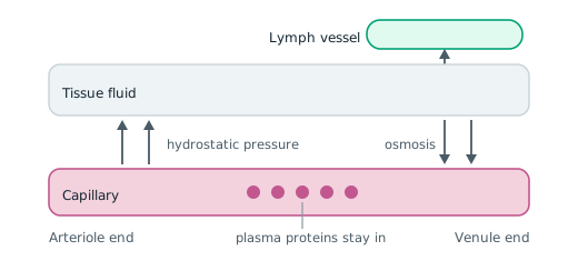
Read the figure from left to right, because the two ends of one capillary are doing opposite things. The arrows point out on the left and back in on the right, and nothing about the capillary itself has changed in between. What changed is which force is winning.
The plasma proteins are the part to notice. They are drawn inside the capillary at both ends, and they never leave. That is why the blood's water potential keeps falling as water is lost, and it is why water comes back at all. Take the proteins out of the figure and there is no return arrow.
The lymph vessel is the admission that the two arrows do not balance. More water leaves than returns, so the leftover has to go somewhere, and the vessel drains it back to the blood further along.
This balance can be upset. A low concentration of plasma proteins raises the blood's water potential, so less water returns by osmosis and fluid builds up. High blood pressure raises the hydrostatic pressure, forcing out more fluid than can be reabsorbed. A blockage in the lymphatic system stops excess fluid draining. In each case, fluid accumulates in the tissues.
Cardiovascular risk factors
A risk factor is an aspect of a person's lifestyle, or a substance in their body or environment, that has been shown to be linked to a higher rate of disease. For cardiovascular disease, risk factors include age, a diet high in salt or saturated fat, smoking, a lack of exercise, and genetics. As always, a risk factor is a correlation with disease, and does not on its own prove that it directly causes it.