AQA A-Level Biology: The Human Gas Exchange System
A clear revision guide to the human gas exchange system for AQA A-Level Biology: the lungs, alveoli, how we breathe in and out, and how lung disease affects gas exchange.
Humans are large, active and warm-blooded, so we need to take in a lot of oxygen quickly. This guide covers the structure of the lungs, why the alveoli are such good exchange surfaces, the mechanism of breathing, and what happens when disease disrupts it.
The structure of the gas exchange system
Air travels a branching path into the lungs. It passes down the trachea (windpipe), which divides into two bronchi, one to each lung. Each bronchus branches into many smaller bronchioles, which end in clusters of tiny air sacs called alveoli. Each alveolus is wrapped in a dense network of capillaries, so that air and blood are brought very close together.
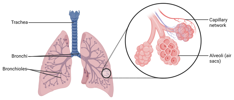
Follow the branching and the reason for it becomes obvious. One tube becomes two, two become many, and the many end in hundreds of millions of alveoli. A single wide tube into each lung would carry the same air, but it would offer almost no surface for that air to be absorbed through. The branching is how a volume gets turned into an area.
Why alveoli are excellent exchange surfaces
The alveoli show every feature of a good exchange surface. Their walls, the alveolar epithelium, are just one flattened cell thick, giving a very short diffusion distance, and there are hundreds of millions of alveoli, giving an enormous surface area. The surface is moist, so gases dissolve before diffusing, and permeable to oxygen and carbon dioxide. The dense capillary network keeps blood flowing past, which maintains the concentration gradient.
When you breathe in, oxygen diffuses from the air in the alveolus, across the alveolar epithelium and the capillary wall, into the blood, down its concentration gradient. Carbon dioxide diffuses the other way, from the blood into the alveolus, to be breathed out.
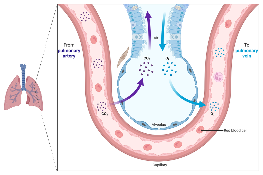
Three things on that diagram are worth reading carefully, because each is a mark.
The two gases move in opposite directions at the same time, and each moves down its own concentration gradient. Oxygen is more concentrated in the alveolar air than in the blood arriving from the body, so it diffuses in. Carbon dioxide is more concentrated in that blood than in the air, so it diffuses out. Neither is pumped and neither follows the other.
The blood arrives from the pulmonary artery and leaves in the pulmonary vein. This is the one place in the body where the artery carries deoxygenated blood and the vein carries oxygenated blood, which is worth fixing now because questions exploit it.
Breathing is what keeps the gradients steep. Air moving in and out replaces the alveolar air, so the oxygen concentration inside stays high and the carbon dioxide concentration stays low. Blood flowing past does the same from the other side. Stop either and diffusion slows as the two sides equalise.
Ventilation: breathing in and out
Ventilation is the movement of air in and out of the lungs. It matters because it constantly refreshes the air in the alveoli, bringing in air rich in oxygen and removing air high in carbon dioxide, which maintains the concentration gradients across the alveolar surface.
Breathing works by changing the volume, and therefore the pressure, of the chest (thoracic) cavity. Air always moves from high pressure to low pressure.
Breathing in (inspiration) is active. The diaphragm contracts and flattens, and the external intercostal muscles contract to pull the ribcage up and out (the internal intercostals relax). This increases the volume of the thoracic cavity and so lowers the pressure inside it below atmospheric pressure, and air flows into the lungs down the pressure gradient.
Breathing out (expiration) at rest is passive. The diaphragm and external intercostal muscles relax, the ribcage drops, and the elastic tissue in the alveoli recoils. This decreases the volume of the thoracic cavity and raises the pressure above atmospheric, so air flows out. Because the elastic recoil does the work, the internal intercostal muscles do not usually need to contract at rest; they mainly contract during forceful breathing out, such as during exercise.
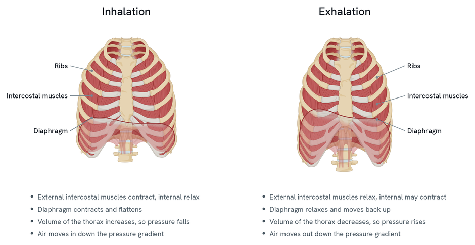
Compare the two ribcages rather than reading them one at a time. The whole of ventilation is in the difference: on the left the diaphragm is flat and the ribcage is wide and high, on the right the diaphragm is domed and the ribcage lower and narrower. Everything else follows from that change in volume.
The external and internal intercostal muscles are described as antagonistic because they pull the ribcage in opposite directions.
One thing the diagram cannot show, and which is worth adding: air is not sucked or pushed. The muscles change only the volume of the thorax. Pressure then changes because the same air occupies a different space, and air moves down the pressure gradient that results. An answer that says the lungs pull air in has skipped the step the marks are for.
How lung disease reduces gas exchange
Diseases interfere with either the exchange surface or the ability to ventilate.
Some reduce the rate of gas exchange directly. Fibrosis thickens the alveolar tissue, increasing the diffusion distance. Emphysema breaks down the walls between alveoli, reducing the surface area. Both, along with a loss of elasticity, lower the rate at which oxygen can enter the blood.
Others affect ventilation. Loss of elasticity, as in fibrosis, means the lungs cannot expand or recoil as well, reducing the volume of each breath (the tidal volume) and the total that can be forced out (the forced vital capacity). Asthma inflames and narrows the bronchi, restricting airflow and reducing the volume that can be breathed out in one second (the forced expiratory volume).
Because less oxygen reaches the blood, a person with lung disease often breathes faster to compensate. Even so, their cells receive less oxygen, so the rate of aerobic respiration falls, less ATP is made, and they feel fatigued.
Reading the evidence: correlation and causation
Lung disease is linked to risk factors such as smoking and air pollution. When you look at data on these, it is important to separate a correlation from a causal relationship. A correlation means that a change in one variable is matched by a change in another, which you can see on a scatter diagram. Causation means one variable actually causes the change in the other. A correlation on its own does not prove causation, because a third, hidden factor may be responsible for both. Establishing cause needs careful, controlled investigation, not just an association.