AQA GCSE Biology: The Brain
A Triple award GCSE Biology revision guide to the brain: the cerebral cortex, cerebellum and medulla, how brain function is mapped, and why the brain is hard to study and treat.
This whole guide is Triple award only. The brain is the control centre of the nervous system, made of billions of neurones. This guide covers the main regions of the brain and what they do, how scientists work out which region does what, and why the brain is so difficult to study and to treat. The theme is that the brain's complexity and delicacy make it hard to investigate.
The regions of the brain
The brain has several regions, each with its own job.
- Cerebral cortex: the highly folded outer layer, responsible for consciousness, intelligence, memory and language.
- Cerebellum: at the back, below the cerebral cortex, responsible for muscle coordination, movement and balance.
- Medulla: at the base, connecting to the spinal cord, controlling unconscious activities such as heartbeat and breathing rate.
A useful way to keep the three apart is to ask what would be lost if each were damaged. Lose the cerebral cortex and you lose thinking, memory and speech. Lose the cerebellum and you stay conscious and able to think, but your movements become clumsy and unbalanced. Lose the medulla and breathing and heartbeat stop, which is why damage there is fatal even though it controls nothing you are ever aware of.
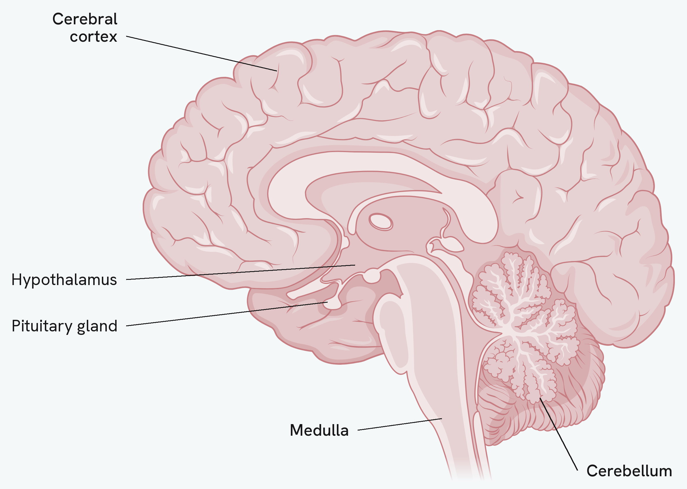
The diagram also labels two structures you meet elsewhere in this topic: the hypothalamus, which monitors and controls body temperature, and the pituitary gland, the master gland of the endocrine system. Both are worth knowing the position of, but only the cortex, cerebellum and medulla are examined as brain regions here.
Mapping brain function
(Higher tier only.) Scientists have worked out what different regions do in three main ways.
- Studying patients with brain damage: seeing which abilities are lost when a particular region is damaged reveals that region's function.
- Electrical stimulation: applying a small current to a specific area and watching the effect on the body or behaviour.
- MRI scanning: watching which regions become most active during different tasks.
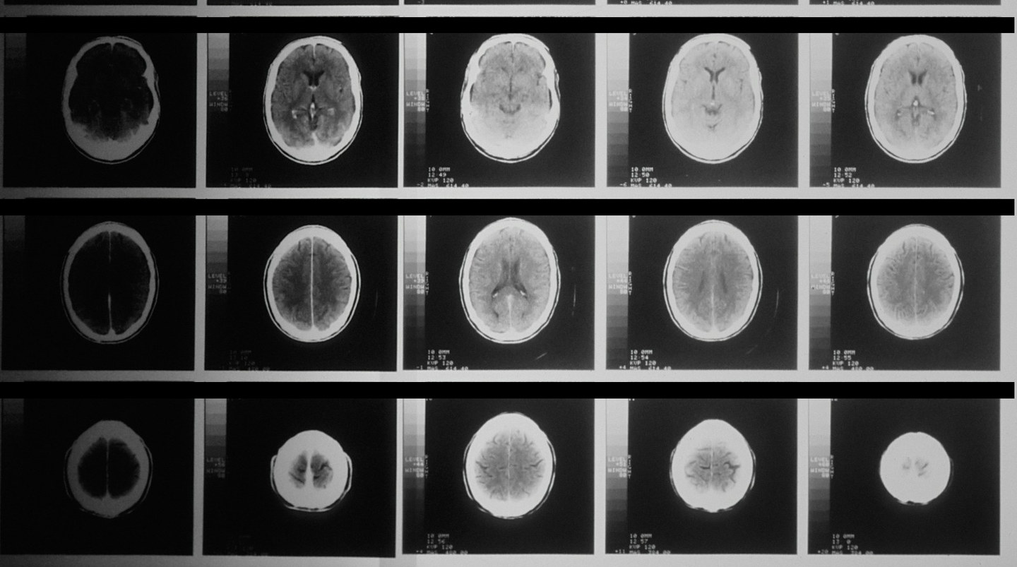
These are CT scans: a stack of cross-sections through one head, each taken a centimetre higher than the last. A scan like this shows the brain's structure, which is what lets damage be found without surgery, so it is what makes the first method in the list possible. MRI is the one named in the specification because it can also show which regions become active while someone carries out a task.
Why the brain is hard to study and treat
(Higher tier only.) Several features of the brain make it difficult to investigate and to treat.
- It is extremely delicate, so surgery risks damaging areas other than the one being treated.
- It is highly complex, with billions of interconnected neurones, so it is hard to work out exactly which region is responsible for a given function.
- It is encased in the skull, so reaching it at all means major surgery.
- The membranes surrounding the brain stop many drugs passing from the blood into brain tissue, so treatments that would work elsewhere in the body cannot get to it.
Each of the three research methods has a matching weakness, and questions often ask you to evaluate rather than list them. Brain damage studies rely on whatever injuries happen to occur, so scientists cannot choose which region is damaged or control anything else about the patient. Electrical stimulation requires the skull to be opened, so it is usually only possible during surgery carried out for another reason. MRI scanning shows which regions are active during a task, but activity is not the same as responsibility, and several regions light up at once.
There is an ethical dimension too. The brain is the organ most tied to personality and identity, so the risk of causing permanent harm, and the question of whether a patient with brain damage can meaningfully consent, both weigh more heavily than they would for research on another organ.
How this fits together
The brain processes the information the senses collect. One of the most important sense organs, and the other Triple-award detail in this section, is the eye, covered in the eye guide.