AQA GCSE Biology: The Eye
A Triple award GCSE Biology revision guide to the eye: its structures, the pupil reflex, accommodation, and the correction of short and long sight.
This whole guide is Triple award only. The eye is the sense organ that detects light. This guide covers the parts of the eye and their jobs, how the pupil reflex adjusts to different light levels, how the lens focuses on near and distant objects, and how short and long sight are corrected. The theme throughout is bending light to focus it on the retina.
The structures of the eye
The eye has several parts, each with a role in detecting and focusing light.
- Retina: contains light-sensitive receptor cells that detect light intensity and colour, and send impulses to the optic nerve.
- Optic nerve: carries the impulses from the retina to the brain.
- Sclera: the tough white outer layer that protects the eye.
- Cornea: the transparent, curved front layer that refracts (bends) light into the eye.
- Lens: a flexible, transparent disc that focuses light onto the retina.
- Iris: a ring of muscle that controls the size of the pupil, regulating how much light enters.
- Ciliary muscles and suspensory ligaments: together these control the shape of the lens.
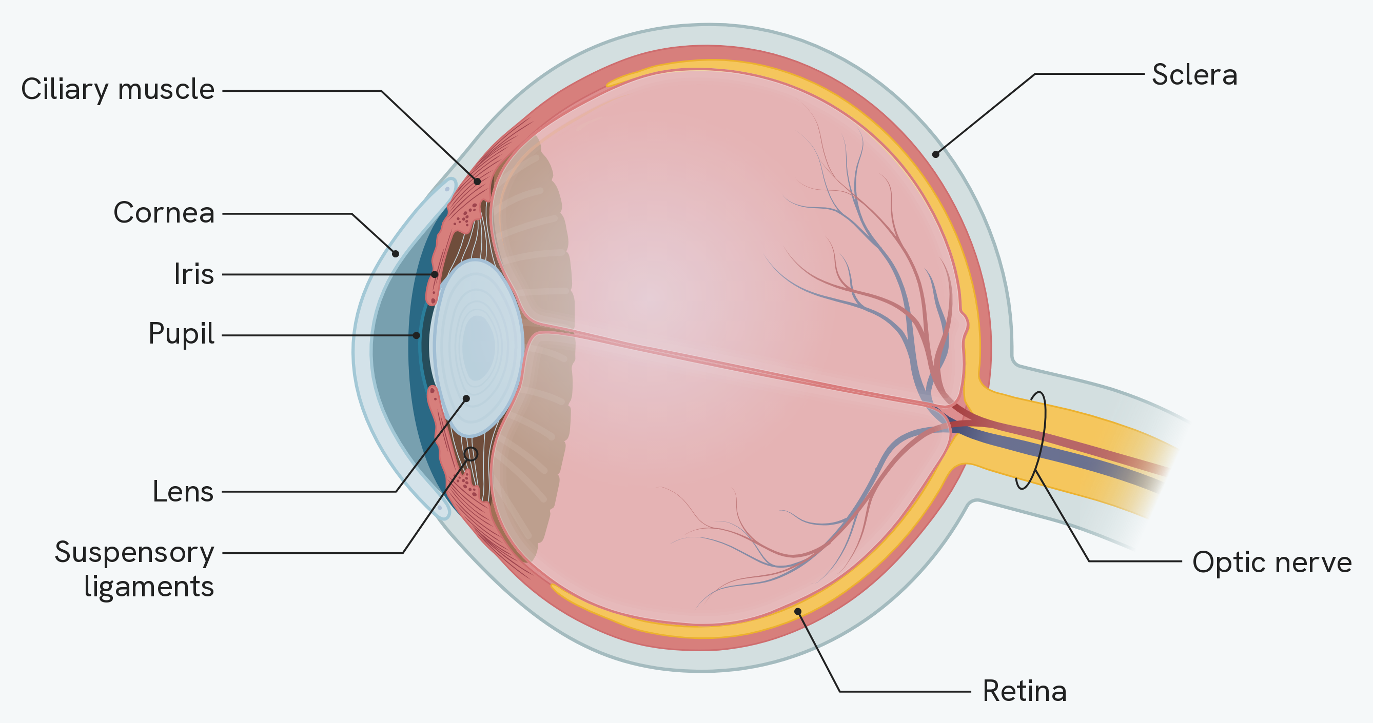
Note the order light travels in: cornea, then pupil, then lens, then retina. The cornea does most of the refraction and the lens fine-tunes the focus, which is why only the lens needs to change shape.
The pupil reflex
The iris adjusts the pupil automatically to control how much light enters.
- In dim light, the pupil dilates (widens): the radial muscles contract and the circular muscles relax, letting more light in.
- In bright light, the pupil constricts (narrows): the circular muscles contract and the radial muscles relax, letting less light in and protecting the retina.
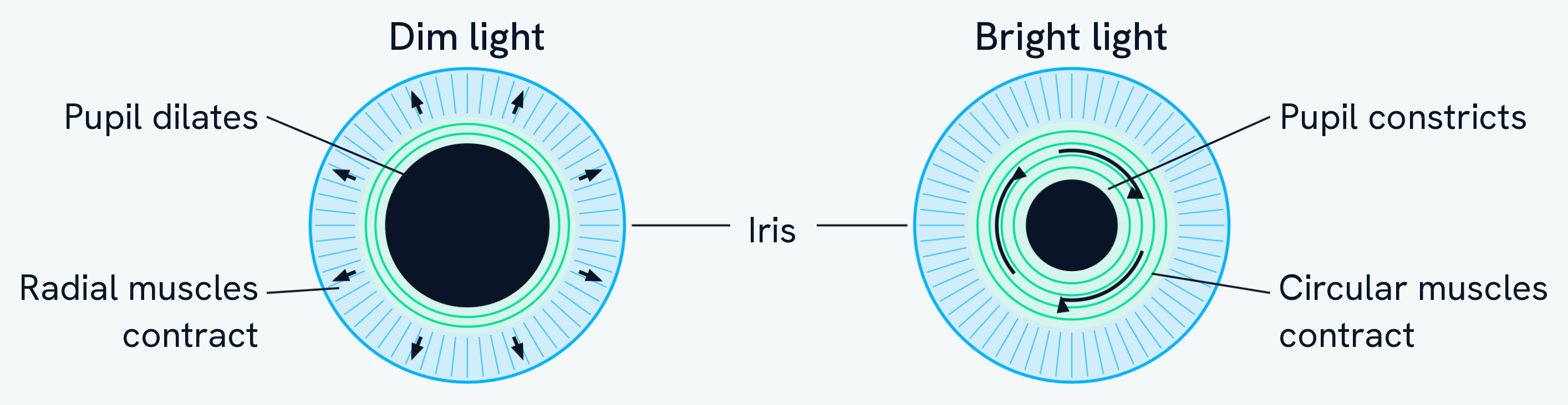
The two sets of muscles pull in different directions, which is the easiest way to remember them. The radial muscles run outwards like spokes, so contracting them pulls the opening wider; the circular muscles run around the pupil like a drawstring, so contracting them pulls it smaller.
Accommodation
Accommodation is how the eye focuses on objects at different distances by changing the shape of the lens.
- To focus on a near object, the ciliary muscles contract, the suspensory ligaments loosen, and the lens becomes thicker (more curved), so it refracts the light more strongly.
- To focus on a distant object, the ciliary muscles relax, the suspensory ligaments pull tight, and the lens is pulled thin (flatter), so it refracts the light less strongly.
In both cases, the aim is to focus the light exactly on the retina.
Eye defects and their correction
Two common defects happen when light is not focused on the retina.
- Myopia (short-sightedness): distant objects cannot be focused, because light focuses in front of the retina (the eyeball is too long or the lens too thick). It is corrected with a concave lens, which spreads the light out before it enters the eye.
- Hyperopia (long-sightedness): near objects cannot be focused, because light focuses behind the retina (the eyeball is too short or the lens too thin). It is corrected with a convex lens, which brings the light together more before it enters the eye.
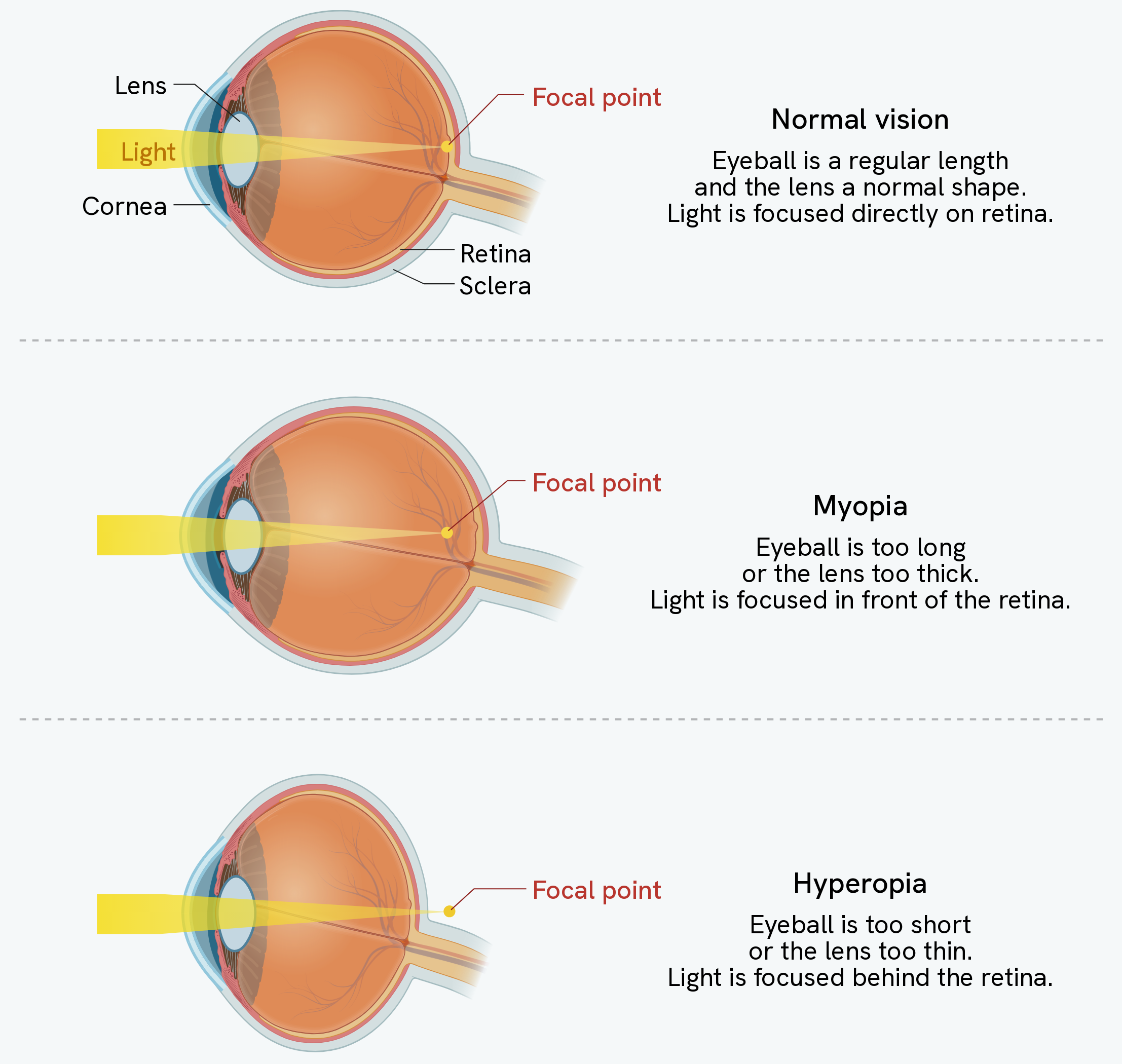
Compare where the yellow focal point sits in each eye. That single position is what the whole topic turns on: on the retina is normal, in front of it is myopia, behind it is hyperopia.
Note that each defect has two possible causes, and the diagram gives both. The eyeball being the wrong length is the one students usually write, but a lens that is too thick or too thin produces exactly the same result, and the mark scheme accepts either.
These defects can also be treated with contact lenses, laser eye surgery (which reshapes the cornea) or replacement lens surgery (an artificial lens is inserted).
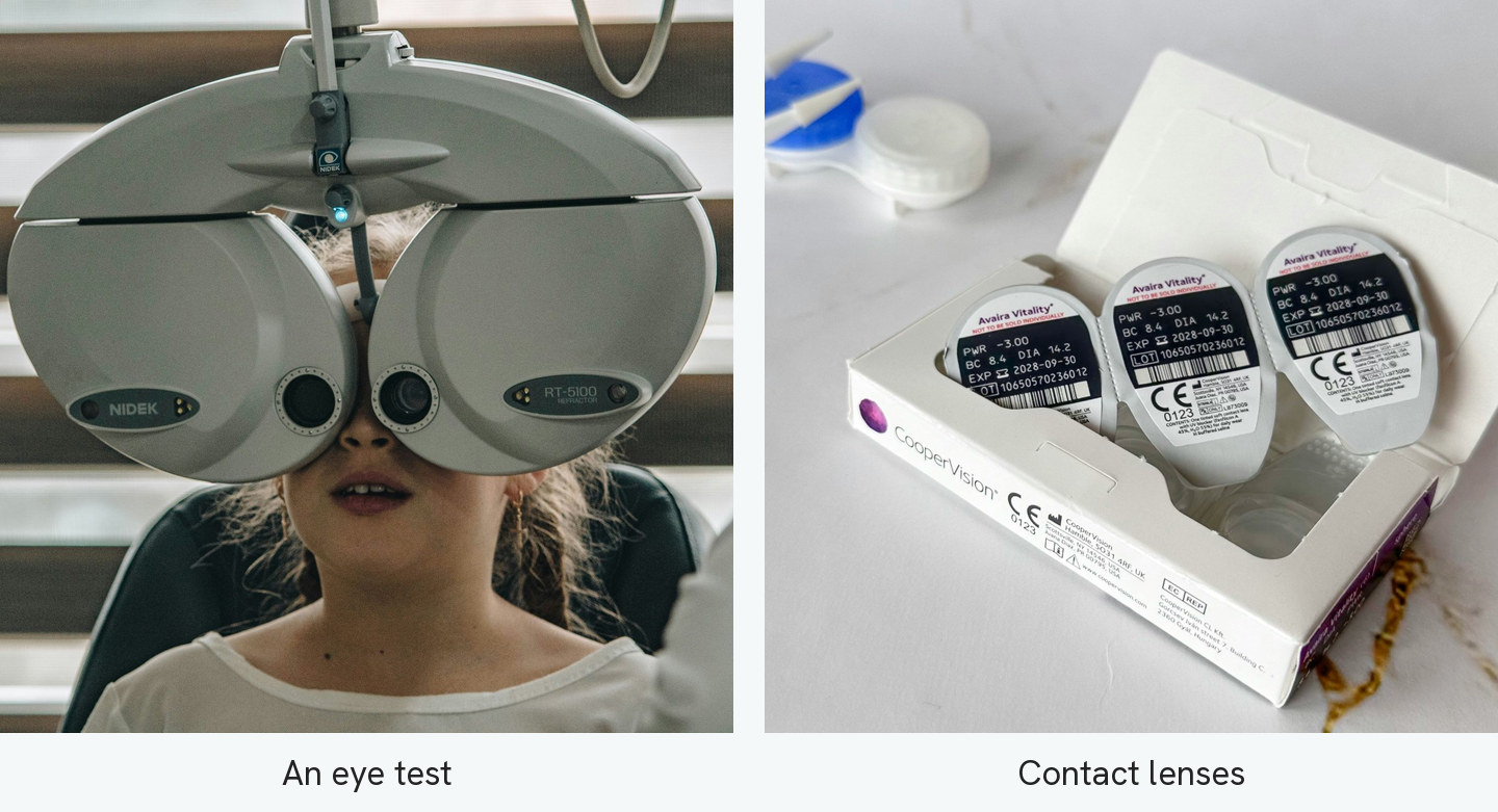
The instrument on the left holds lenses of different powers in front of each eye in turn until the letters come into focus, which measures how much refraction the eye is failing to do for itself. The packets on the right are printed PWR −3.00. A negative power means a concave lens, so these are lenses for myopia — the correction described above, made in a form that sits on the cornea instead of in front of it.
How this fits together
The eye and brain work together to sense the world. Another Triple-award part of coordinating the body is keeping its temperature steady, covered in the control of body temperature guide.