AQA GCSE Biology: Microscopy
A clear GCSE Biology revision guide to microscopy: magnification and resolution, light and electron microscopes, the magnification formula, and converting units.
Cells are far too small to see with the naked eye, so we use microscopes. This guide explains the two things a microscope does, magnify and resolve, the difference between light and electron microscopes, and how to use the magnification formula, including the unit conversions that so often catch people out. The maths here is worth practising until it is automatic.
Magnification and resolution
A microscope does two jobs, and they are not the same thing.
- Magnification is how many times larger the image appears compared with the real object.
- Resolution is the shortest distance between two points that can still be seen as separate. It sets the level of detail you can see.
A blurry image made bigger is still blurry, so high resolution matters as much as high magnification.
The clearest way to feel the difference is a car at night. From far away its two headlights look like a single blob of light. Walk towards it and at some point the blob splits into two separate lights. Nothing got bigger at that moment; your eye simply reached the distance at which it could resolve them. Magnification would have given you a larger blob. Only better resolution gives you two headlights.
This is exactly why a photograph enlarged too far goes fuzzy rather than detailed, and it is the answer to any question asking why magnification alone is not enough.
Light and electron microscopes
There are two main types.
- A light microscope forms an image using visible light.
- An electron microscope forms an image using beams of electrons.
Electron microscopes have much higher magnification and much higher resolution than light microscopes. This lets scientists study cells in far finer detail and see smaller sub-cellular structures, such as ribosomes, that a light microscope cannot resolve.
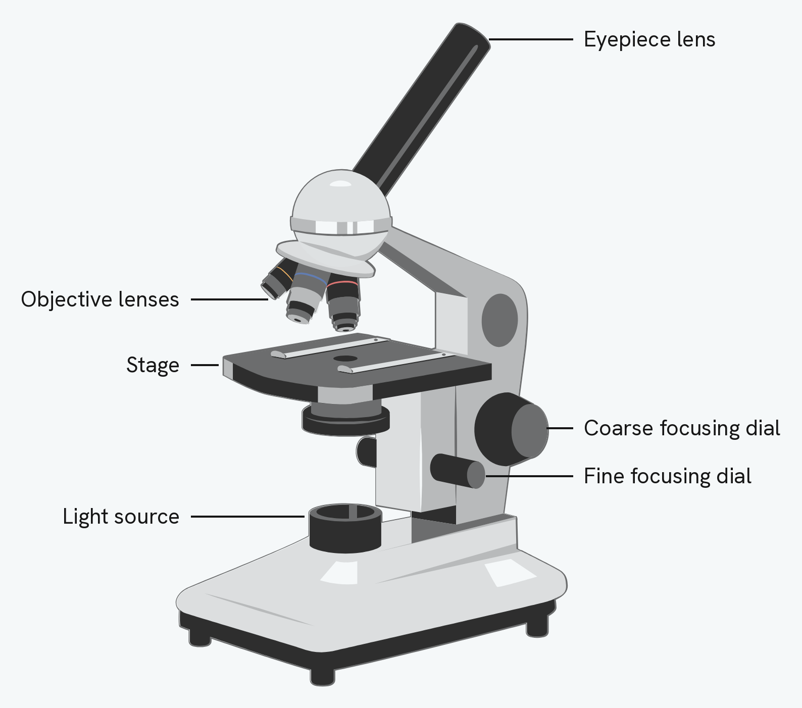
AQA likes to ask how electron microscopes changed our understanding of cells, which is a question about history rather than equipment. Structures such as ribosomes, and the internal detail of mitochondria and chloroplasts, were simply invisible before electron microscopes existed. Sub-cellular structures were not discovered by looking harder down a light microscope; they were discovered when a tool arrived that could resolve them.
Light microscopes are still used, and it is worth being able to say why. They are cheaper, easier to use, and can show living specimens, which an electron microscope cannot.
The magnification formula
Magnification links the size of the image to the real size of the object:
image size = actual size × magnification
This can be rearranged:
- magnification = image size ÷ actual size
- actual size = image size ÷ magnification
The single most important rule is to make sure the image size and actual size are in the same unit before you calculate.
Magnification is a ratio, so it has no units. The answer is written as "×1500", never "1500 μm". Writing a unit after a magnification loses the mark.
Converting units
Cell sizes are measured in millimetres (mm), micrometres (μm) and sometimes nanometres (nm), and you often have to convert between them.
- To go from a larger unit to a smaller one, multiply by 1000 at each step (mm → μm is ×1000).
- To go from a smaller unit to a larger one, divide by 1000 at each step (μm → mm is ÷1000).
For example, a structure with an actual length of 30 μm magnified 40 times gives an image of 30 × 40 = 1200 μm, which is 1200 ÷ 1000 = 1.2 mm.
Work the other way too, because that is the more common exam version. If a drawing of a cell measures 50 mm across and the magnification is ×2000, the actual size is 50 ÷ 2000 = 0.025 mm, which is 0.025 × 1000 = 25 μm. A real cell is only ever a few micrometres across, so an answer in whole millimetres is a sign you have divided the wrong way round.
You may also be asked to give an answer in standard form. That same 25 μm is 2.5 × 10⁻⁵ m, and marks are available for writing it correctly.
How this fits together
Microscopy is how we see the cells and structures from the earlier guides. Microscopes also let us watch cells divide, which is part of the cell cycle covered in the chromosomes and the cell cycle guide.