AQA A-Level Biology: Control of Blood Glucose
A clear revision guide to blood glucose control for AQA A-Level Biology: insulin, glucagon and adrenaline, glycogenesis and gluconeogenesis, the second messenger model, and types I and II diabetes.
The concentration of glucose in the blood has to be held steady: too little starves the cells of respiratory fuel, and too much draws water out of them by osmosis. This guide explains how the pancreas detects and corrects changes in blood glucose using the hormones insulin and glucagon, how adrenaline and the second messenger model fit in, and what goes wrong in diabetes. It is a clear example of hormonal negative feedback.
Why blood glucose must be controlled
Glucose is the main respiratory substrate, so its level in the blood must be kept within limits.
- If it falls too low, there is not enough glucose for respiration, especially in the brain and nervous system, so too little ATP is made and processes such as active transport fail.
- If it rises too high, the blood has a lower water potential, so water moves out of cells and tissues into the blood by osmosis. The kidneys also cannot reabsorb all the glucose, so glucose and extra water are lost in the urine, causing dehydration.
Blood glucose rises after eating carbohydrates, which are digested and absorbed into the blood, and falls during exercise, which increases the rate at which glucose is respired.
Insulin lowers blood glucose
Insulin is secreted by the beta cells of the islets of Langerhans in the pancreas when blood glucose is too high. It lowers blood glucose by acting on the liver and muscle cells.
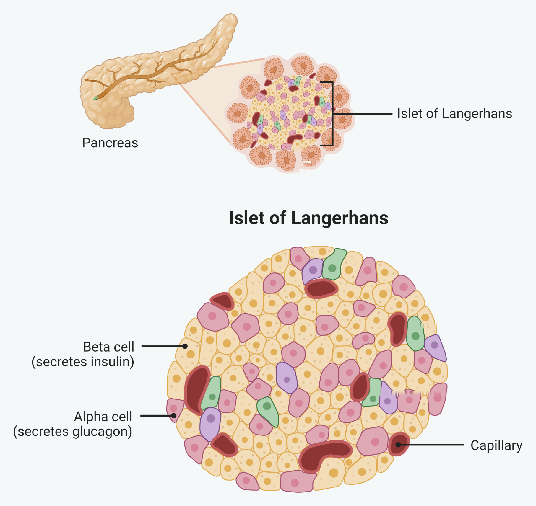
The two cell types to know are on that figure. Beta cells secrete insulin and alpha cells secrete glucagon, and both sit in the same islet, so the pancreas can raise or lower blood glucose from the same place.
Notice the capillaries running through the islet. That is how a hormone secreted by a cell in the pancreas reaches a liver cell: straight into the blood, and from there to every cell in the body.
- Insulin binds to receptors on the cell surface membranes of liver and muscle cells.
- This increases the number of glucose channel proteins in the membrane, so cells take up more glucose from the blood by facilitated diffusion.
- It activates enzymes that convert glucose into glycogen for storage, a process called glycogenesis.
- It also increases the rate of respiration of glucose.
Together these remove glucose from the blood, bringing the level back down.
Glucagon and adrenaline raise blood glucose
Glucagon is secreted by the alpha cells of the islets of Langerhans when blood glucose is too low. It acts mainly on the liver.
- Glucagon binds to receptors on liver cells.
- It activates enzymes that break glycogen back down into glucose, a process called glycogenolysis.
- It activates enzymes that make new glucose from glycerol and amino acids, a process called gluconeogenesis.
- It also decreases the rate of respiration of glucose.
Adrenaline, from the adrenal glands, also raises blood glucose, and is released when glucose is low or when the body is stressed or exercising. It binds to liver cell receptors, activates glycogenolysis, inhibits glycogenesis, and stimulates the secretion of glucagon, all of which release more glucose for respiration.
Because insulin and glucagon have opposite effects and are released under opposite conditions, they form a negative feedback loop that holds blood glucose steady.
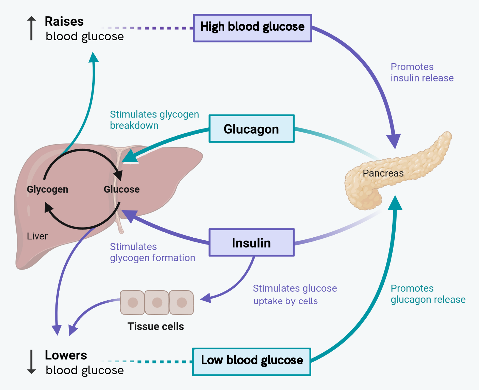
Read it as one loop, not two halves. Follow it clockwise from the top and the pattern is the same each time: a change is detected, the pancreas releases a hormone, the liver responds, and the change is reversed. That reversal is what makes it negative feedback.
The liver sits at the centre because of the two arrows between glycogen and glucose. The same store is built up by insulin and broken down by glucagon, and which way the arrow runs is the whole of this topic.
The second messenger model
Adrenaline and glucagon do not enter the cell; they trigger changes from the outside using a second messenger.
- The hormone binds to a specific receptor on the cell surface membrane.
- This activates the enzyme adenylate cyclase inside the membrane.
- Adenylate cyclase converts ATP into cyclic AMP (cAMP), the second messenger.
- The cAMP activates protein kinase A, which sets off a cascade of reactions that break glycogen down into glucose.
- The glucose then leaves the liver cell by facilitated diffusion into the blood.
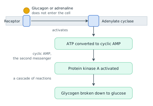
Everything in this model follows from the hormone staying outside. It is a protein, so it cannot cross the phospholipid bilayer, and yet the glycogen it needs broken down is inside. The whole chain exists to get a message across a membrane the messenger cannot pass.
That is also where the name comes from. The hormone is the first messenger, and it gets no further than the receptor. Cyclic AMP is the second messenger, and it is the one that actually carries the signal through the cytoplasm. If a question asks what the second messenger is, the answer is cAMP, never the hormone.
The order of the four names is the other thing worth having cold, because questions ask for the sequence: receptor, then adenylate cyclase, then cyclic AMP, then protein kinase A. Two of those are in the membrane and two are in the cytoplasm, which is a useful way to keep them in order.
The advantage of this model is amplification: a single hormone molecule causes many molecules of cAMP to be made, and each of those activates many enzymes, so the signal is greatly magnified and glucose is released rapidly.
Diabetes
Diabetes is a condition in which blood glucose cannot be controlled properly, so it rises too high and stays high, especially after meals. There are two types.
- In type I diabetes, the beta cells produce too little or no insulin. It usually develops in childhood, when an autoimmune response destroys the beta cells. It is controlled with insulin injections, matched to glucose intake by monitoring blood glucose with biosensors, together with eating regularly and managing carbohydrate intake to avoid sudden rises. Insulin cannot be taken as a tablet because it is a protein and would be digested by protease enzymes in the gut.
- In type II diabetes, the pancreas still makes insulin, but the receptors on the target cells lose their sensitivity to it, so cells take up less glucose. It is linked to obesity, and it is managed by controlling the diet (less sugar and fat, and foods with a low glycaemic index), taking regular exercise, losing weight, and sometimes drugs that make the receptors more responsive.
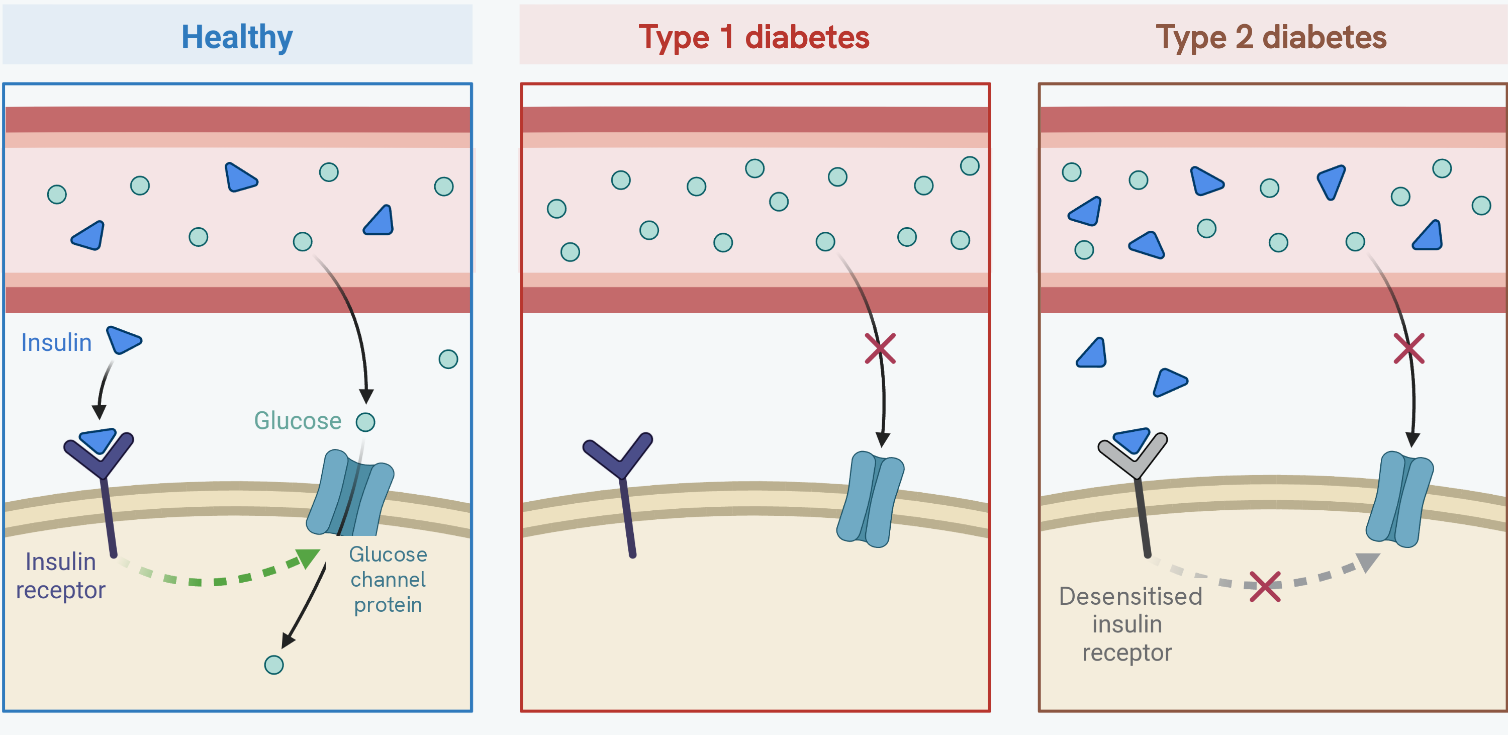
Compare the three panels at one point: what is in the blood. In the healthy panel and in type 2 there is insulin in the blood; in type 1 there is none. That single difference is what separates the two types, and it is why type 1 is treated with injected insulin and type 2 is not.
Then look at the receptor. In type 2 the insulin is there and it does bind, but the receptor has lost its sensitivity, so nothing happens downstream and the glucose channel proteins never reach the membrane. Writing that a person with type 2 diabetes makes no insulin is the common error, and this figure is the answer to it.
Glucose can be measured to help diagnose and monitor diabetes. A known glucose solution can be used with Benedict's test and a colorimeter to build a calibration curve, and an unknown sample is then compared against it. Glucose appears in the urine of someone with high blood glucose because not all of it can be reabsorbed in the kidney, as the glucose carrier proteins become saturated and work at their maximum rate.
How this fits together
Blood glucose control is hormonal negative feedback: the pancreas senses the level and releases insulin or glucagon to correct it, amplified where needed by the second messenger model. The kidney appeared here in passing, as the organ that reabsorbs glucose and adjusts water loss, and its full role in filtering the blood and balancing water is covered in the kidney and osmoregulation guide.