AQA A-Level Biology: Muscles and Contraction
A clear revision guide to muscles for AQA A-Level Biology: antagonistic pairs, the structure of a sarcomere, the sliding filament theory of contraction, and slow and fast twitch muscle fibres.
Muscles are the effectors that let animals move. This guide explains why muscles have to work in pairs, how a muscle is built from overlapping protein filaments, and how those filaments slide over each other to make the muscle shorten. It ends with the two types of muscle fibre and why each suits a different kind of activity. The central idea is the sliding filament theory, and everything else supports it.
Antagonistic pairs
A muscle can only pull, by contracting; it cannot push itself back to its original length. So muscles are arranged in antagonistic pairs, where one muscle reverses the movement caused by the other.
- The muscle that contracts to produce a movement is the agonist; it pulls on a bone.
- The other muscle, the antagonist, relaxes.
To make the opposite movement, the roles swap. In the arm, the biceps and triceps form such a pair: the biceps contracts to bend the arm while the triceps relaxes, and the triceps contracts to straighten it while the biceps relaxes. Working together, a pair can also hold a joint steady and maintain posture.
Muscles are joined to bone by tendons, while ligaments join bone to bone. Skeletal muscle is incompressible, which means it can transmit the full force of its contraction to the bone rather than simply squashing.
The structure of a muscle
A muscle is made of bundles of muscle fibres, which are the muscle cells, packed together. These cells are specialised.
- Their cell membrane is called the sarcolemma and their cytoplasm the sarcoplasm.
- They contain many myofibrils, the contractile threads, made of two proteins: actin (thin filaments) and myosin (thick filaments).
- They have many nuclei and a great deal of endoplasmic reticulum (here called the sarcoplasmic reticulum), along with many mitochondria to supply ATP.
Each myofibril is divided into repeating units called sarcomeres, and it is the sarcomere that actually contracts.
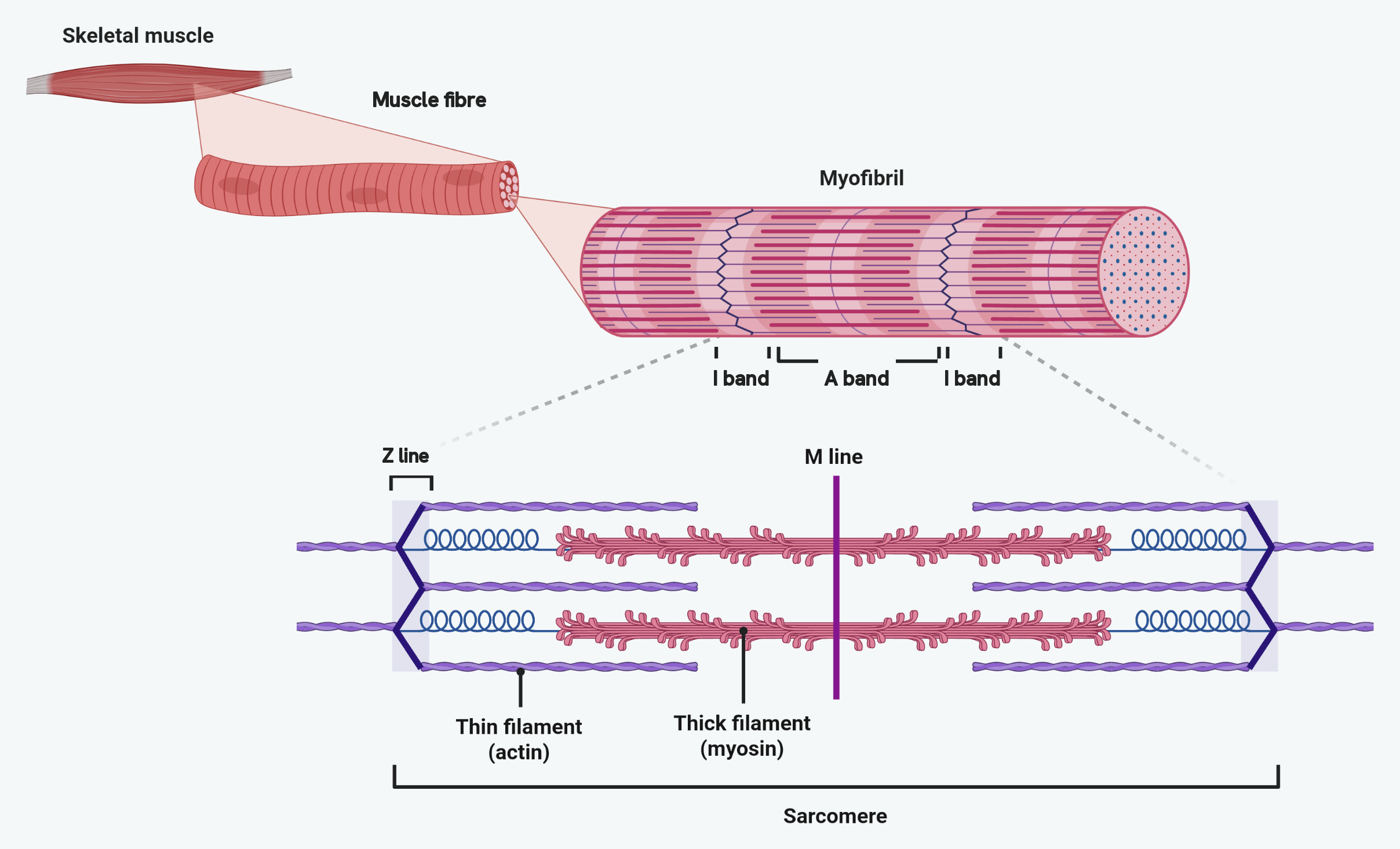
Read it as four magnifications of the same thing, not four different things. A muscle is a bundle of muscle fibres; each fibre is packed with myofibrils; each myofibril is a chain of sarcomeres; and each sarcomere is actin and myosin, overlapping.
The striped look of the myofibril comes from that overlap, which is where the bands get their names. Where there is actin alone the myofibril is pale, and that is the I band. Where the myosin lies, whether or not actin overlaps it, it is dark, and that is the A band.
The sarcomere
A sarcomere is made of partly overlapping myosin and actin filaments, and it has a regular pattern of bands.
| Feature | What it is |
|---|---|
| Z-line | Marks the two ends of a sarcomere |
| M-line | The middle of the sarcomere |
| H-zone | The central region containing only myosin |
| I-band | A light band containing only actin |
| A-band | A dark band spanning the full length of the myosin, including where actin overlaps it |
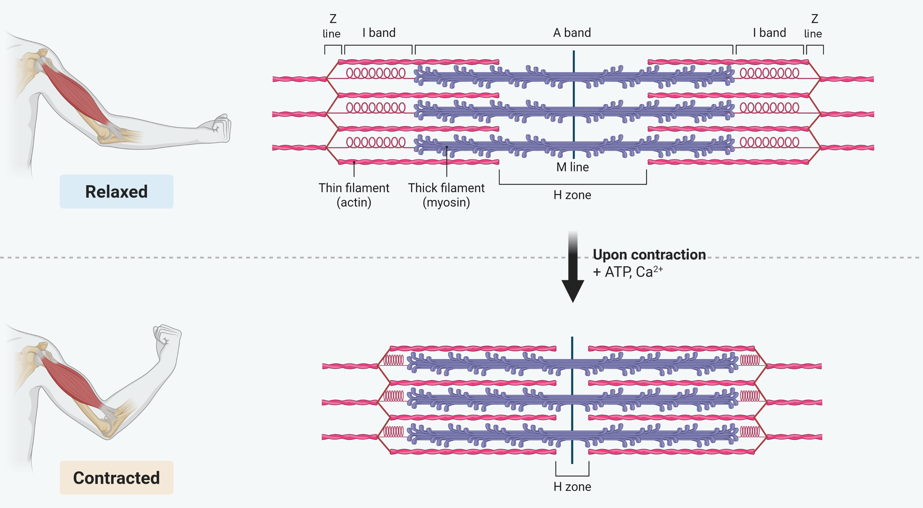
When a muscle contracts, the filaments slide further past each other, so the sarcomere gets shorter. The bands change in a way that tells you which filaments have moved: the H-zone gets shorter, the I-band gets shorter, the Z-lines move closer together, but the A-band stays the same length, because the myosin filaments themselves do not change length.
Compare the two halves of that figure band by band, because that comparison is the exam question. Three things get narrower and one does not, and the one that does not is the A band. Nothing has shortened: the actin has simply slid further in over the myosin, and the myosin is as long as it ever was.
The H zone is the part worth checking carefully. It is the stretch of myosin with no actin overlapping it, so as the actin slides inwards the H zone is squeezed from both sides. If a question asks why it narrows, the answer is the overlap, not the length of any filament.
The sliding filament theory
Contraction happens because the myosin heads pull the actin filaments inwards, sliding them over the myosin so the sarcomere shortens. When many sarcomeres do this at once, the whole muscle contracts. The process runs in a repeating cycle.
- An action potential spreads down the muscle fibre and along infoldings called T-tubules, causing the sarcoplasmic reticulum to release calcium ions into the sarcoplasm.
- The calcium ions cause tropomyosin, which has been blocking the binding sites on the actin, to move away and expose the binding sites.
- Myosin heads attach to the exposed sites on actin, forming actinomyosin cross-bridges.
- The myosin heads pivot, pulling the actin along, then a molecule of ATP binds to each head, making it detach from the actin.
- The ATP is hydrolysed by the enzyme ATPase, which is activated by the calcium ions, releasing energy that returns the myosin head to its original position.
- The head then binds further along the actin and the cycle repeats, pulling the filaments past each other, for as long as calcium ions and ATP are present.
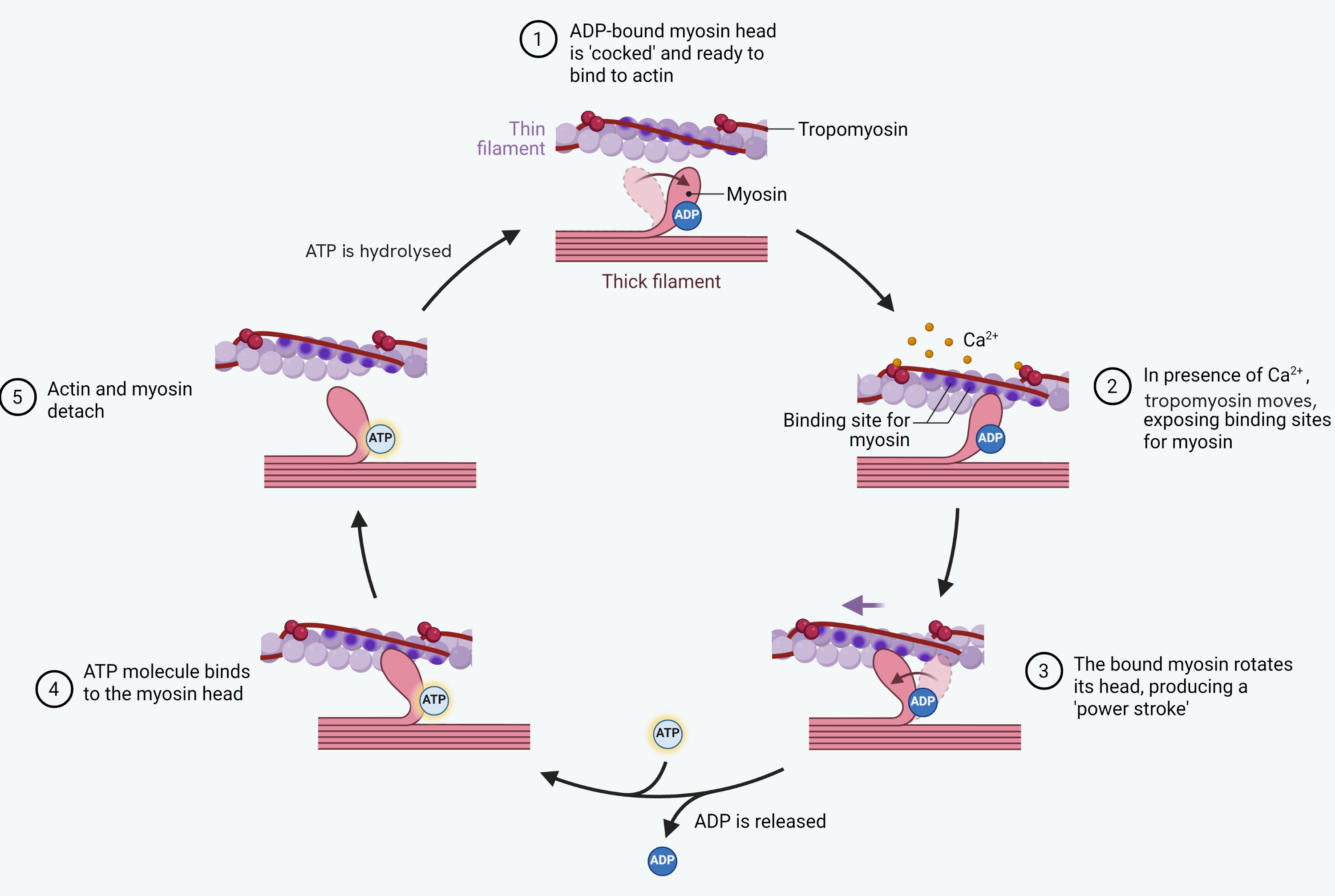
Go round the loop once and note where the ATP appears, because that is what most questions are really asking. ATP binds at stage four to make the head let go, and it is hydrolysed on the way back round to re-cock the head. Both uses are worth marks, and the one students leave out is the first: without ATP the head cannot detach at all.
Watch the thin filament at the top of the loop as well. The dark strand running along it is tropomyosin, and in the first stage it lies across the binding sites so no myosin head can attach. Calcium ions move it off them, and that is the whole of the trigger: no calcium, no exposed sites, no contraction.
When stimulation stops, the muscle relaxes. Calcium ions are actively transported back into the sarcoplasmic reticulum using ATP, tropomyosin moves back to block the binding sites again, and with no cross-bridges able to form the filaments slide back.
Slow and fast twitch fibres
Muscles contain two types of fibre, suited to different demands.
| Feature | Slow twitch | Fast twitch |
|---|---|---|
| Specialised for | Slow, sustained contraction (endurance) | Rapid, powerful contraction in short bursts |
| Example | Postural muscles, long-distance runners' legs | Sprinters' leg muscles |
| Respiration | Aerobic, releasing energy slowly | Anaerobic, releasing energy quickly |
| Myoglobin | High, giving a red colour and an oxygen store | Low, giving a pale colour |
| Mitochondria | Many | Few |
| Blood supply | Many capillaries | Fewer capillaries |
| Glycogen | Less | Much, for glycolysis |
Slow twitch fibres are built for endurance. Their many mitochondria, rich blood supply and high myoglobin (which stores oxygen) all support a high rate of aerobic respiration, and their good blood supply also removes lactate before it builds up and causes fatigue.
Fast twitch fibres are built for power. They rely on anaerobic respiration in the cytoplasm, with plenty of stored glycogen to fuel glycolysis and a high concentration of the enzymes it needs. Because glycolysis makes only two ATP per glucose and produces lactate, these fibres tire quickly. They also store phosphocreatine, which rapidly regenerates ATP from ADP by donating a phosphate group. Phosphocreatine works anaerobically without producing lactate, but it runs out within a few seconds, so it only powers short, intense bursts of effort.
How this fits together
The sliding filament theory ties the whole topic together: a nerve impulse crosses the neuromuscular junction, calcium ions are released, and myosin heads walk the actin filaments inwards using ATP. Muscle is one kind of effector at the end of the response pathway. Keeping that pathway, and the rest of the body, working within stable limits is the job of homeostasis, covered in the homeostasis and control of heart rate guide.