AQA A-Level Biology: The Kidney and Osmoregulation
A clear revision guide to the kidney for AQA A-Level Biology: the nephron, ultrafiltration, selective reabsorption, the loop of Henle as a counter-current multiplier, and the control of water by ADH.
The kidneys filter the blood, keeping what the body needs and removing waste and excess water. This guide follows the blood through a nephron, the kidney's working unit: first the filtering step of ultrafiltration, then the selective reabsorption of useful substances, then the clever water-saving trick of the loop of Henle, and finally how the hormone ADH fine-tunes water loss. The recurring theme is control of water potential.
The kidney and the nephron
Blood enters each kidney through the renal artery, carrying excess water and urea, and leaves through the renal vein, now filtered and carrying less water and urea. Any excess water and urea removed leaves as urine, which passes down the ureter to the bladder.
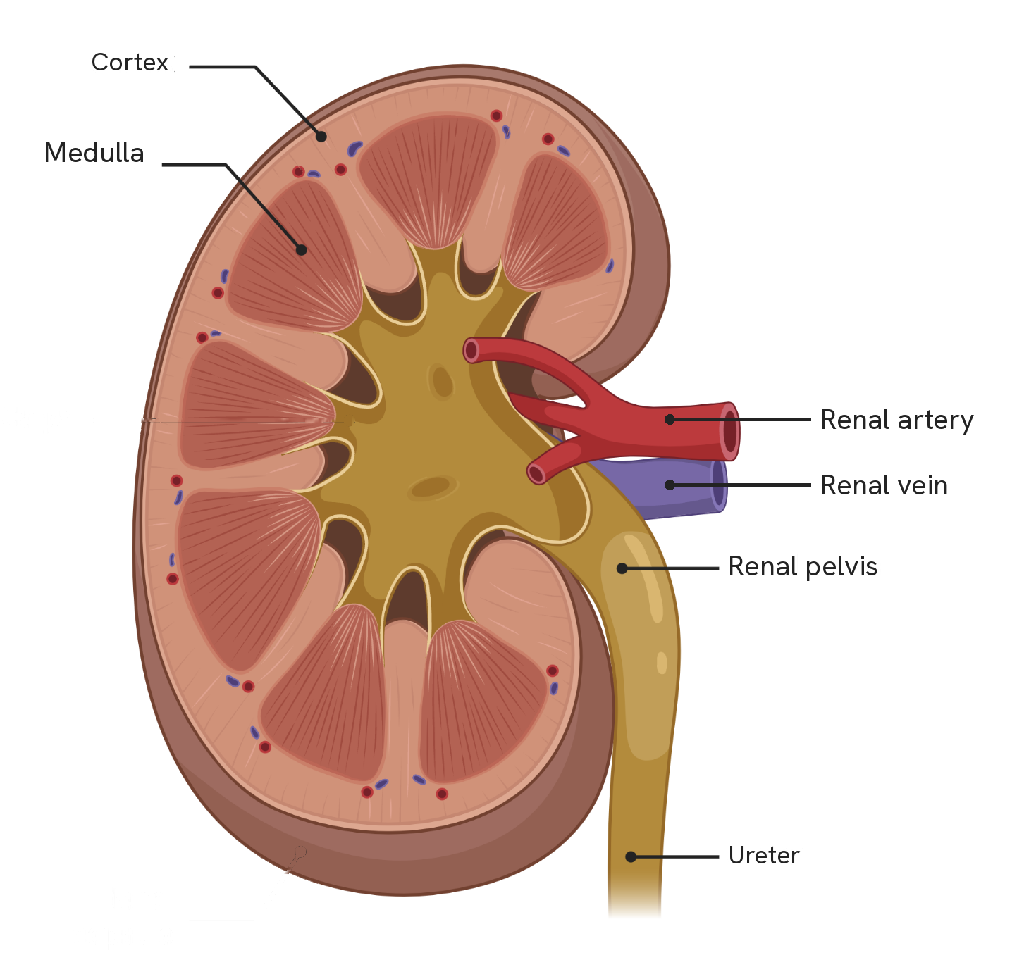
Six labels, and no more than six: the cortex, the medulla, the renal artery, the renal vein, the renal pelvis and the ureter. Diagrams of the kidney elsewhere often add a calyx and a renal capsule, which are not on this specification and are not worth the time.
The two vessels are the pair to be sure of, because they are what a question will give you. The renal artery carries blood in, with the urea and excess water still in it; the renal vein carries blood out, with less of both. Everything the rest of this guide describes happens between those two labels.
Note also where the cortex and medulla sit relative to each other, because the nephron runs through both. The Bowman's capsule and the convoluted tubules are in the cortex; the loop of Henle dips down into the medulla and back. A question asking which part of the kidney a structure is found in is asking you to read this figure.
The filtering is done by around a million nephrons, the functional units of the kidney, where urine is made. Blood is delivered to each nephron by the afferent arteriole and taken away by the efferent arteriole. Urine is made in two main stages: ultrafiltration and selective reabsorption.
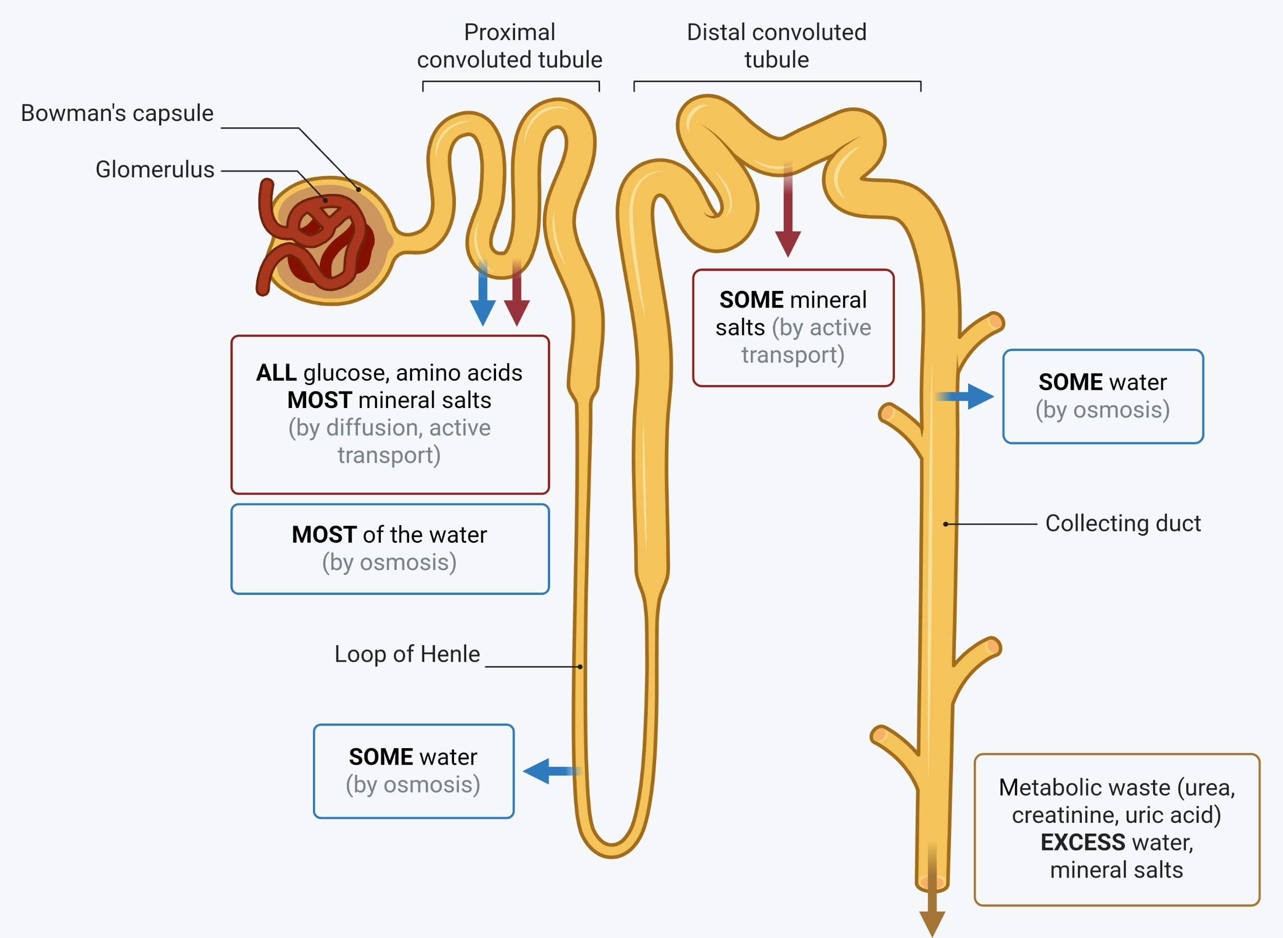
Learn the parts in the order the filtrate meets them, because questions give you a stage and ask what happens there. Bowman's capsule, then proximal convoluted tubule, then loop of Henle, then distal convoluted tubule, then collecting duct.
The words on the figure are doing real work. ALL the glucose and amino acids are taken back in the proximal convoluted tubule, and the word is all: glucose in the urine means the reabsorption has failed, not that some was meant to be left. Everywhere else the word is most or some, and whatever is not taken back carries on down the tubule.
Ultrafiltration
Ultrafiltration happens between the glomerulus, a knot of capillaries, and the Bowman's capsule that surrounds it. It is driven by high pressure.
Because the efferent arteriole is narrower than the afferent arteriole, blood entering the glomerulus is squeezed into a smaller space, which builds up a high hydrostatic pressure. The blood has also arrived almost directly from the heart, so it is already at high pressure. This pressure forces small molecules out of the blood and into the Bowman's capsule.
The fluid passes through three layers, each adapted to filter.
- The capillary endothelium is one cell thick and has pores (fenestrations) that let fluid through.
- The basement membrane is a fine mesh of proteins that acts as a filter, letting small molecules pass but holding back large ones.
- The podocytes lining the capsule have finger-like projections that leave filtration slits between them.
Small, soluble molecules, such as water, glucose, amino acids, salts, urea and hormones, pass into the capsule to form the glomerular filtrate. Large proteins, such as albumin and antibodies, and blood cells are too big to pass and stay in the blood.
Selective reabsorption
The filtrate contains useful substances that must not be lost, and these are taken back into the blood in the proximal convoluted tubule (PCT). This is where all the glucose and amino acids, and most of the salts and water, are reabsorbed.
The PCT is adapted for this. It is highly folded (tightly wound) and its cells have microvilli and many mitochondria, all of which support active reabsorption over a large surface area. Reabsorption works by a mechanism that depends on sodium.
- Sodium ions are actively transported out of the tubule cells into the blood, lowering the sodium concentration inside the cells.
- Sodium ions then move from the filtrate into the cells by facilitated diffusion, co-transporting glucose and amino acids in with them.
- The glucose and amino acids pass on into the blood by facilitated diffusion.
- Removing all these solutes into the blood lowers its water potential, so water follows by osmosis, and is reabsorbed too.
Some substances are not reabsorbed. Urea, being small and lipid-soluble, partly diffuses back into the blood, but wastes such as uric acid and creatinine are not reabsorbed because there are no transport proteins for them and they cannot cross the membrane freely, so they stay in the filtrate to be excreted.
The loop of Henle
The loop of Henle lets the kidney reabsorb water and produce urine more concentrated than the blood. It works as a counter-current multiplier, building up a high concentration of sodium ions deep in the medulla, which lowers the water potential there.
- The ascending limb is impermeable to water. Sodium ions are actively transported out of it into the medulla. Because water cannot follow, this raises the sodium concentration of the medulla and lowers its water potential.
- The descending limb is permeable to water. As the filtrate passes through the low water potential of the medulla, water leaves it by osmosis into the surrounding tissue, where it is carried away by capillaries, so the filtrate becomes more concentrated as it descends.
- This arrangement of fluid flowing in opposite directions in the two limbs multiplies the effect, so a very low water potential builds up towards the bottom of the loop.
The importance of this is felt later: the low water potential of the medulla means that as filtrate passes through the collecting duct, water can be drawn out of it by osmosis. A longer loop of Henle creates a lower water potential and lets more water be reabsorbed, which is why desert animals have very long loops.
Osmoregulation and ADH
Osmoregulation is the control of the water and salt content of the body. The final adjustment of water is made in the distal convoluted tubule (DCT) and collecting duct, controlled by the hormone ADH, which changes how permeable their walls are to water.
- When the water potential of the blood is too low (too little water), the hypothalamus detects it and the pituitary gland releases more ADH. ADH makes the walls of the DCT and collecting duct more permeable to water, by inserting more aquaporin channels, so more water is reabsorbed into the blood and a small volume of concentrated urine is produced.
- When the water potential of the blood is too high, less ADH is released, the walls become less permeable, less water is reabsorbed, and a large volume of dilute urine is produced.
This is a negative feedback loop that keeps the water potential of the blood steady. ADH acts through a second messenger system: it binds to receptors on the tubule cells, activating adenylate cyclase to make cyclic AMP, which triggers vesicles carrying aquaporins to fuse with the membrane, opening more water channels.
How this fits together
The kidney is a masterclass in controlling water potential: ultrafiltration by pressure, selective reabsorption driven by sodium co-transport, the counter-current loop of Henle, and fine control by ADH through negative feedback. Balancing water is one of the body's core homeostatic jobs, alongside balancing temperature, which is covered in the temperature regulation guide.