AQA A-Level Biology: Homeostasis and Control of Heart Rate
A clear revision guide to homeostasis for AQA A-Level Biology: negative and positive feedback, why multiple feedback mechanisms are used, and how heart rate is controlled by the SAN, AVN, baroreceptors and chemoreceptors.
Homeostasis is how the body keeps its internal conditions steady while the outside world changes. This guide explains the principle behind it, negative feedback, and why the body uses several feedback mechanisms rather than one. It then works through a detailed example: how the heart sets its own rhythm and how the nervous system speeds it up or slows it down in response to the body's needs. Once you understand negative feedback here, the same logic appears throughout homeostasis.
The principle of homeostasis
Homeostasis is the maintenance of a stable internal environment within narrow limits. It matters because the body's enzymes, and its cells in general, only work well within a small range of temperature, pH and concentration.
The main mechanism is negative feedback: receptors detect when a level has strayed too high or too low, and effectors act to counteract the change and bring the level back to normal. Body temperature control is a good example.
This is different from positive feedback, which amplifies a change and drives a level further from normal. Positive feedback is useful for processes that need to happen quickly and completely once started, such as blood clotting, but it is not used in homeostasis, because it would push conditions further out of balance rather than restoring them.
The body often controls a single factor with several negative feedback mechanisms at once. This gives more precise control, because separate mechanisms can push a level in different directions, actively raising it or lowering it as needed, and it allows a faster, more sensitive response than a single mechanism could.
Control of heart rate
The heart provides a clear example of negative feedback in action. First, though, the heart sets its own basic rhythm.
Cardiac muscle is myogenic, meaning it can contract and relax on its own without signals from nerves. The rhythm is set by a region of the right atrium called the sinoatrial node (SAN), which acts as the heart's pacemaker.
- The SAN sends out regular waves of electrical activity across both atria, making them contract together.
- A layer of non-conducting tissue stops this wave passing straight to the ventricles.
- Instead the wave reaches the atrioventricular node (AVN), which delays it briefly. This delay lets the atria finish contracting and emptying before the ventricles contract.
- The AVN passes the wave to the bundle of His, a strip of conducting tissue that carries it down to the apex (bottom) of the heart, where it spreads through finer Purkyne fibres.
- This makes the ventricles contract from the bottom upwards, which pushes all the blood up and out of the heart.
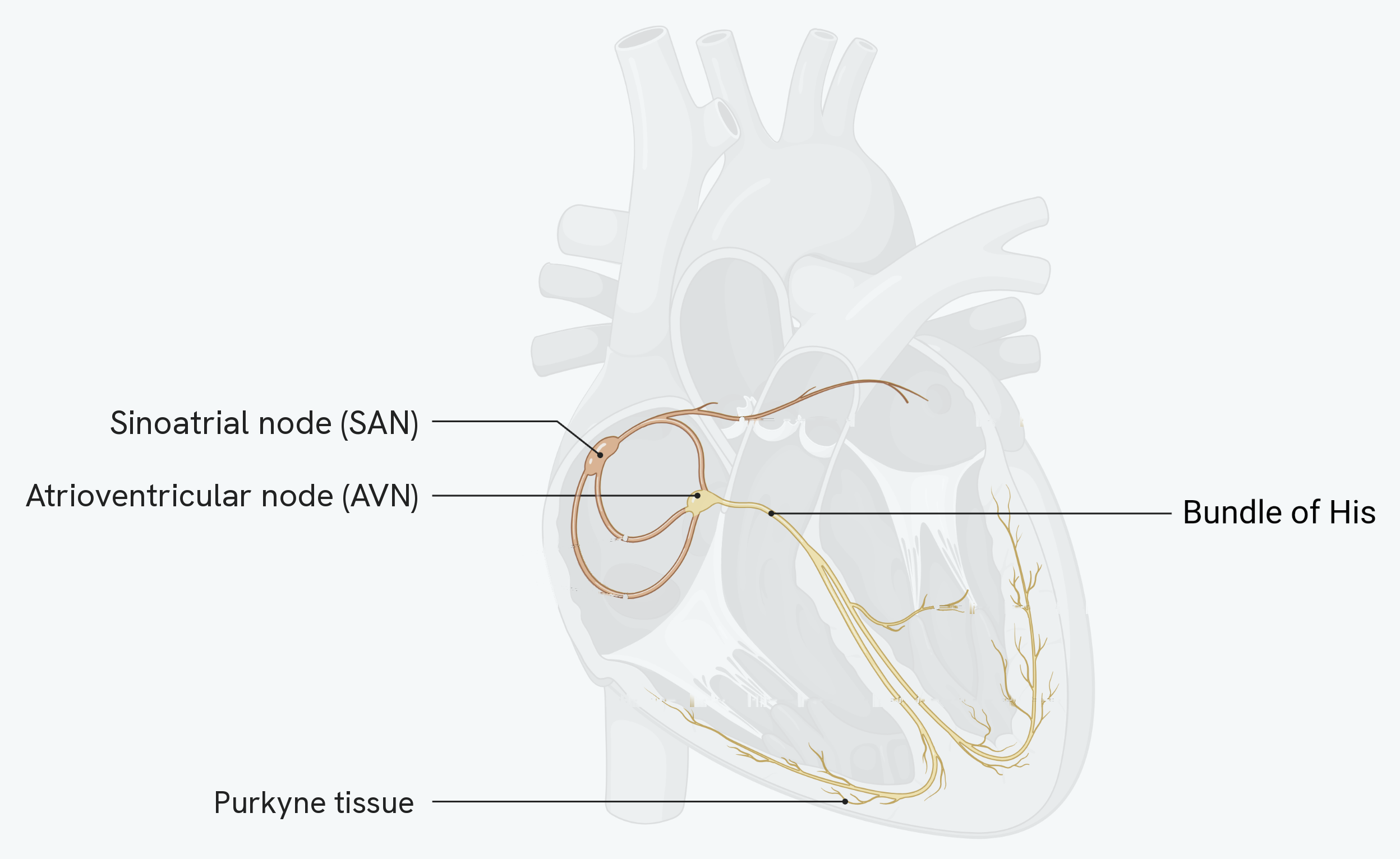
Follow the route in order, because the order is the answer to most questions on this. The wave starts at the SAN in the right atrium, spreads across both atria, is picked up by the AVN, passes down the bundle of His to the apex, and only then spreads back upwards through the Purkyne tissue.
The shape of that path is the point. The bundle of His carries the wave down before the Purkyne tissue carries it up, so the ventricles contract from the bottom upwards and squeeze the blood towards the arteries at the top rather than trapping it at the base.
Adjusting heart rate
The rate of the SAN is adjusted by the medulla oblongata in the brain, using information from two kinds of receptor in the walls of the aorta and carotid arteries.
- Baroreceptors detect blood pressure.
- Chemoreceptors detect the pH, carbon dioxide and oxygen levels of the blood, which change with exercise.
The medulla adjusts the heart through the two branches of the autonomic nervous system: the sympathetic nerve speeds the heart up, and the parasympathetic nerve slows it down. Both work by changing how often the SAN fires.
| Change detected | Response | Effect |
|---|---|---|
| Low blood pressure (baroreceptors) | Impulses sent to the SAN along the sympathetic nerve | Heart rate increases |
| High blood pressure (baroreceptors) | Impulses sent to the SAN along the parasympathetic nerve | Heart rate decreases |
| High CO₂ or low pH (chemoreceptors) | Impulses sent to the SAN along the sympathetic nerve | Heart rate increases |
| Low CO₂ or high pH (chemoreceptors) | Impulses sent to the SAN along the parasympathetic nerve | Heart rate decreases |
Each of these responses restores a healthy level, which is exactly what negative feedback does. During exercise, respiration produces more carbon dioxide, which forms carbonic acid and lowers blood pH; lactate from anaerobic respiration lowers it further. The chemoreceptors detect this, and heart rate rises to remove the excess acid quickly, before it can denature enzymes, and to deliver more oxygen to the respiring muscles. It is important that heart rate falls when blood pressure is too high, because sustained high pressure can damage artery walls, and that it rises when pressure is too low, so that respiring tissues still receive enough oxygenated blood.
How this fits together
The control of heart rate shows the whole logic of homeostasis in one system: a set point, receptors that detect departures from it, a coordinator in the brain, and effectors that correct the change through negative feedback. The same pattern controls the body's other internal conditions, including the tightly regulated level of glucose in the blood, which is the subject of the control of blood glucose guide.