AQA A-Level Biology: Receptors
A clear revision guide to receptors for AQA A-Level Biology: the Pacinian corpuscle and generator potentials, rod and cone cells, retinal convergence, sensitivity and visual acuity.
Receptors are the cells that turn a stimulus into a nervous signal. This guide uses two examples to show how they work: the Pacinian corpuscle, which detects pressure, and the rod and cone cells of the eye, which detect light. Both illustrate the same key ideas, that a receptor responds only to one kind of stimulus, and that a stimulus has to be strong enough to trigger a signal at all.
What a receptor does
A receptor detects a stimulus and is specific to one type of stimulus. When a receptor is stimulated, it produces a small change in the electrical charge across its membrane called a generator potential. If the generator potential is large enough to reach a threshold, it triggers a nerve impulse, an action potential, in a sensory neurone. A weaker stimulus that does not reach threshold produces no impulse at all.
The Pacinian corpuscle
The Pacinian corpuscle is a pressure receptor found deep in the skin, especially in the fingers and feet. Because it responds to mechanical pressure, it is a type of mechanoreceptor.
Its structure is the key to how it works. It is the end of a sensory neurone wrapped in layers of connective tissue separated by a gel. The neurone's membrane contains special stretch-mediated sodium ion channels, which open only when the membrane is stretched.
This structure produces a generator potential as follows:
- Pressure deforms the layers of tissue, which stretches the neurone's membrane and its stretch-mediated sodium ion channels.
- The channels open, and sodium ions diffuse in, making the inside of the neurone less negative. This is depolarisation, and the resulting change is the generator potential.
- A greater pressure deforms the membrane more, so more channels open and more sodium ions enter, giving a larger generator potential.
- If the generator potential reaches the threshold, it triggers an action potential that travels along the sensory neurone.
The Pacinian corpuscle shows two general principles neatly: a receptor responds only to its specific stimulus (here, pressure), and a signal is only sent once the generator potential reaches threshold, an example of the all-or-nothing principle.
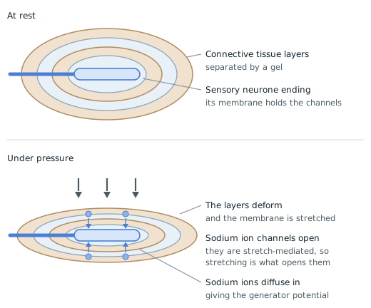
Compare the two panels and only one thing has happened: the layers are flatter. Everything else in the sequence follows from that single mechanical change, which is why this receptor is the one the specification uses to make the point.
The layers are not padding. They are what turns a push on the skin into a stretch of a membrane, and stretch is the only thing these channels respond to. A sodium ion channel that opened in response to pressure directly would not need the onion of tissue around it at all.
Note what the figure does not show: an action potential. What is drawn is sodium ions entering and the membrane becoming less negative, which is the generator potential. Whether that becomes an action potential depends on it reaching the threshold, and a harder press means more channels open, more sodium enters, and a larger generator potential. Calling the thing in this diagram an action potential is the usual way to lose the mark.
Rod and cone cells
The retina contains two kinds of light receptor, or photoreceptor: rod cells and cone cells. They differ in where they are, how sensitive they are, and how much detail they can resolve. Visual acuity means the ability to tell two nearby points apart.
The difference between them comes down largely to how they connect to the neurones that carry signals to the brain, a feature called retinal convergence.
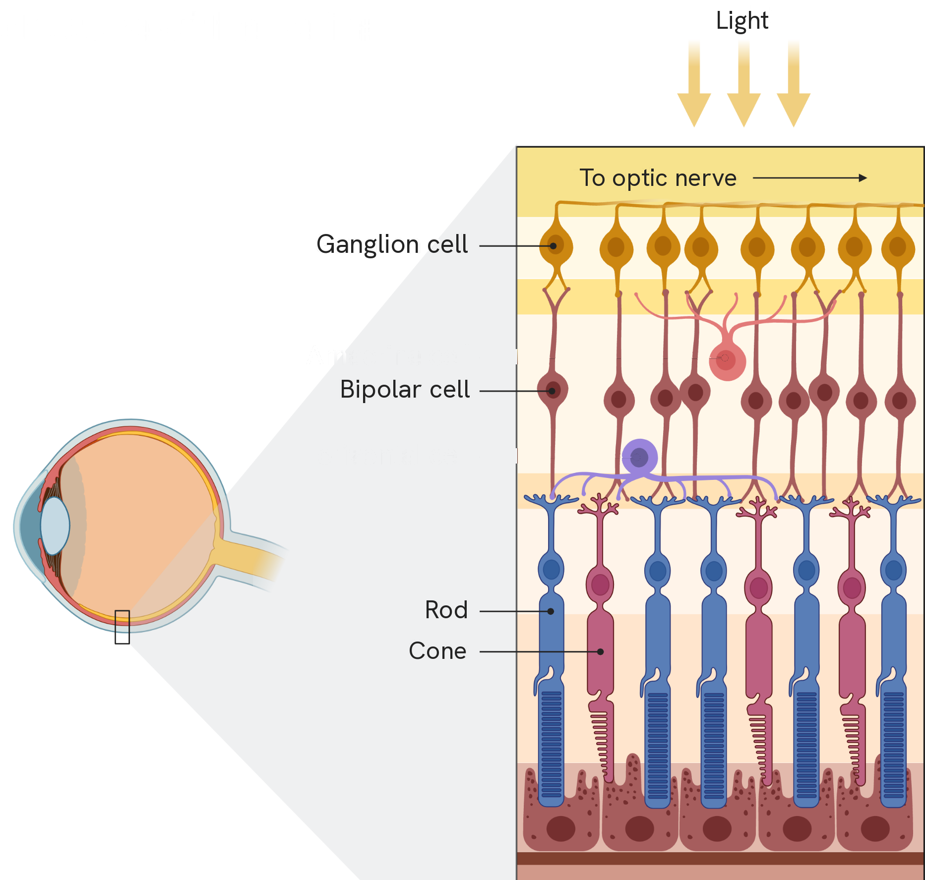
The figure says something about the retina that catches people out: the light reaches the rods and cones last. It passes through the ganglion cells and the bipolar cells first, and the photoreceptors sit at the back, with their light-sensitive ends pointing away from the light.
Follow one rod upwards from the back and you have the path a signal takes: rod or cone, then a bipolar cell, then a ganglion cell, whose fibres leave as the optic nerve. Every difference in the rest of this section is about how many photoreceptors feed into one bipolar cell at that first step.
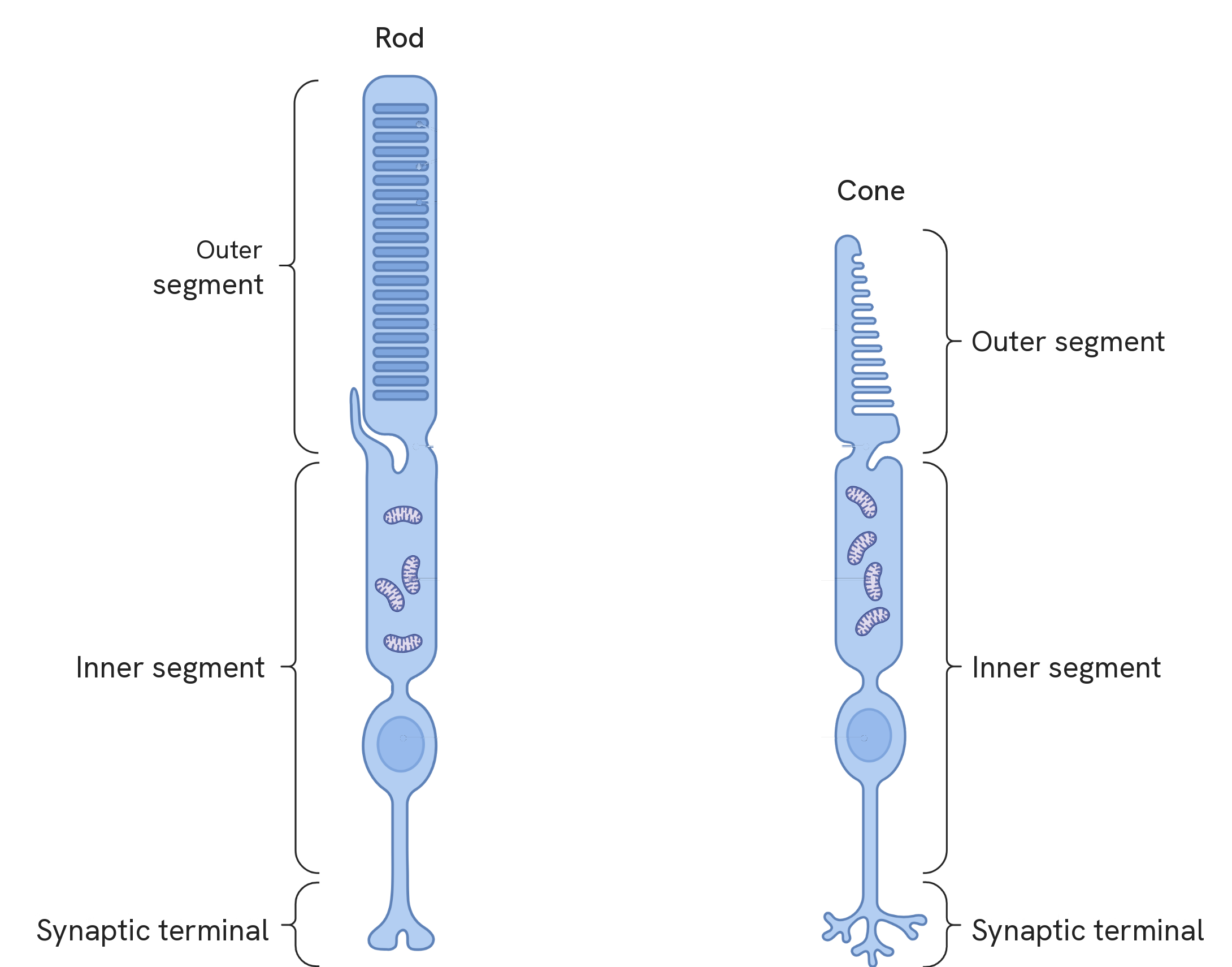
The shapes are where the two names come from: the rod's outer segment is a long, even cylinder, the cone's is short and tapering. That outer segment is the part that holds the pigment, rhodopsin in a rod and iodopsin in a cone, and it is the end that points away from the light.
You are not asked about the inside of either cell. What the figure is useful for is keeping the two apart by sight, and seeing that each one ends in a synaptic terminal, which is where it meets the bipolar cell and where the difference that matters begins.
Rod cells
Rod cells are found in the periphery of the retina and are absent from the fovea. They contain a single pigment, rhodopsin, and there is only one type of rod cell, so they give black-and-white (monochromatic) vision. They are very sensitive to light, so they let us see in dim conditions, but they give low visual acuity.
Both features come from the way rods are wired. Many rod cells connect to a single bipolar cell (and so to a single neurone), a form of retinal convergence.
- This makes them sensitive because of spatial summation: the light hitting one rod alone is not enough to reach threshold, but several rods feeding into the same neurone can add together to reach it, so an impulse is generated even in dim light.
- It also gives them low acuity, because the brain receives just one impulse from the whole group and cannot tell which rod the light came from, so two nearby points are not distinguished.
Cone cells
Cone cells are concentrated in the fovea and are sparse in the periphery. There are three types, each containing a different form of the pigment iodopsin that absorbs red, green or blue light, so cones give colour (trichromatic) vision. They are less sensitive and need bright light, but they give high visual acuity.
Again the wiring explains it. Each cone cell connects to its own neurone.
- This makes them less sensitive, because there is no spatial summation: a single cone must absorb enough light by itself to reach threshold, which only happens in bright conditions.
- It gives high acuity, because two neighbouring cones send two separate impulses to the brain, so two close points can be told apart.
- The three types of cone, absorbing different wavelengths, allow colour vision: stimulating them in different combinations and proportions produces the full range of colours we perceive.
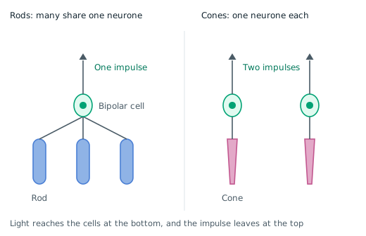
Count the arrows leaving the top of each diagram, because that is all the brain ever receives. Three rods send one. Two cones send two. Every one of the four properties in this section falls out of those two numbers.
On the rod side, three weak signals arrive at the same bipolar cell and add together, which is spatial summation, and it is why dim light is enough to reach threshold. The price is paid in that single arrow: whichever of the three rods the light hit, the brain gets the same one impulse and cannot tell them apart, so two nearby points are seen as one. Sensitive, but low acuity.
On the cone side, nothing adds. Each cone must reach threshold by itself, which takes bright light. What that buys is the second arrow: two cones send two separate impulses, so two nearby points stay two. Less sensitive, but high acuity.
Sensitivity and acuity pull against each other here, and the figure shows why: you cannot add signals together and still know which one came from where.
How this fits together
Both receptors do the same job in the same way: a specific stimulus is converted into a generator potential, and only a stimulus strong enough to reach threshold produces an impulse. What happens to that impulse once it is triggered, how it travels and how it is coordinated, is the subject of the nervous system and neurones guide.